INTRODUCTION
This paper redescribes the Middle Jurassic (Bajocian) foraminifera from Western Australia first described by Frederick Chapman in 1904. This almost-forgotten work (Chapman 1904) illustrated the first Jurassic foraminifera from Australia, and possibly from the Southern Hemisphere, and described three new species and three new varieties of foraminifera, in addition to ostracods which have been redescribed by Malz and Oertli (1993). Because Chapman’s new species names potentially impacted modern identification of Australian Jurassic specimens, it was felt that there was a need to check the validity of Chapman’s names. His collection of specimens, as listed in his paper, were therefore borrowed from the Museum of Victoria, photographed in the ESEM, and described. A list of Chapman’s foraminifera is given in the order in which he described them, on Table 1. Revised identifications are suggested on the same table. The order of species descriptions given here is altered, to correspond with the classification given in Loeblich and Tappan (1987).
Locality of Chapman’s specimens
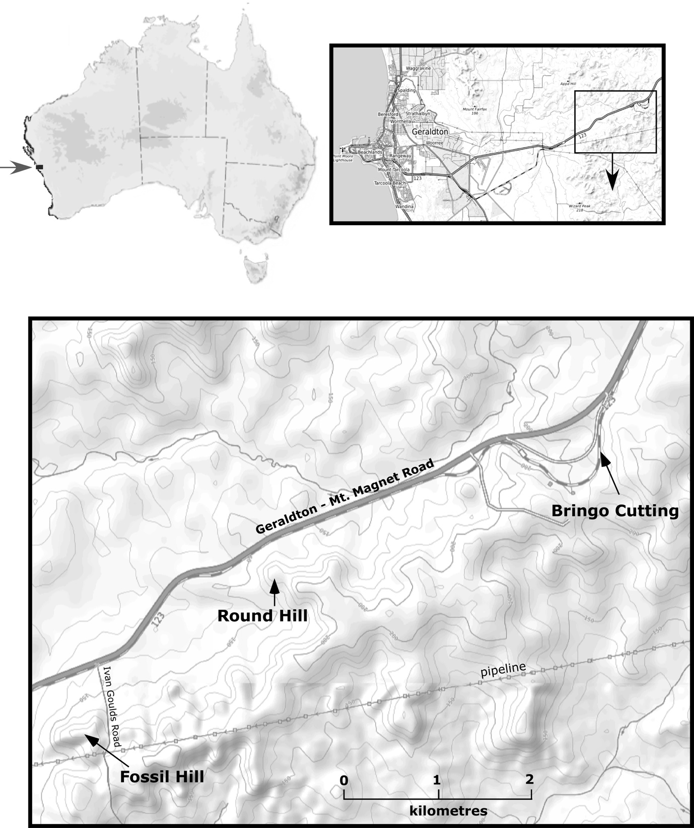
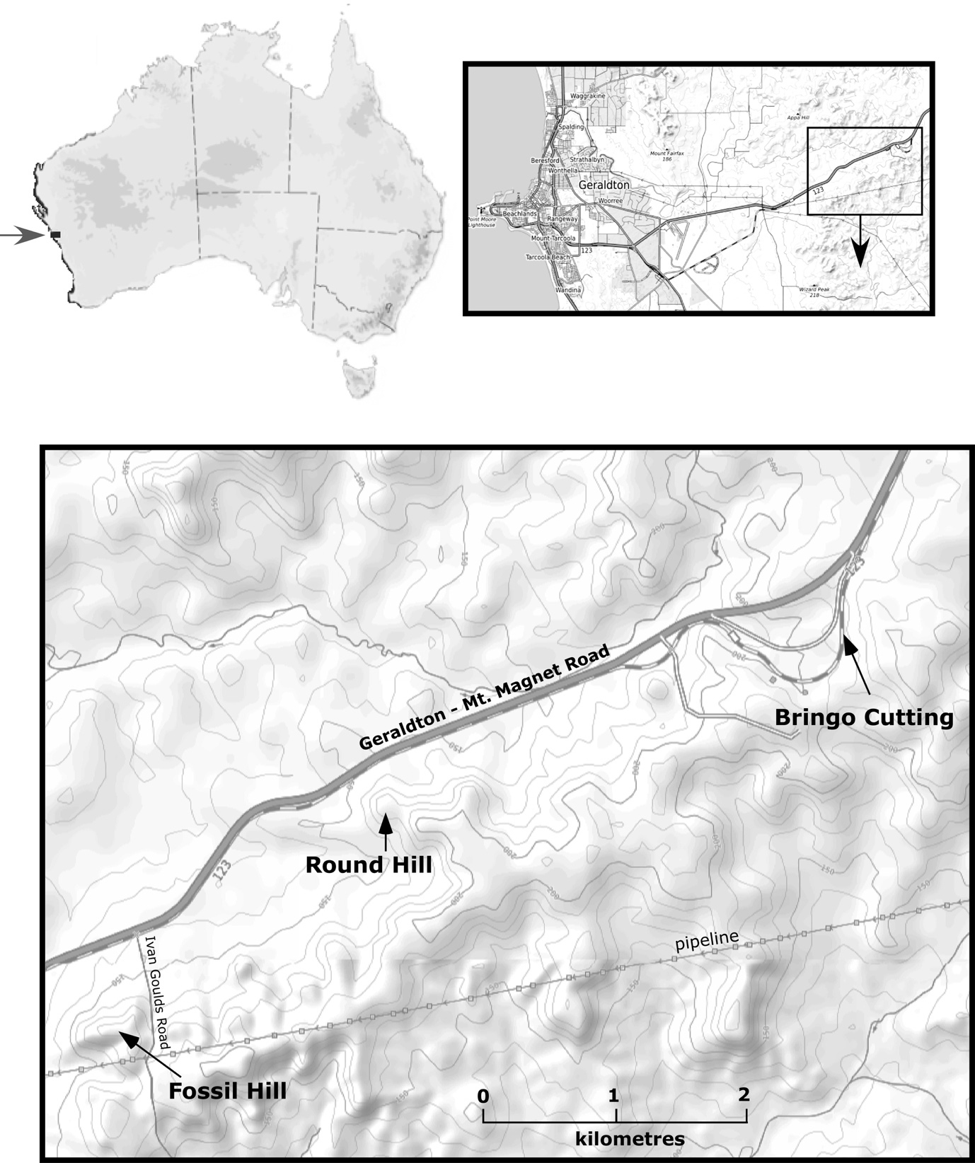
The locality (“near Geraldton, Western Australia”) from which Chapman’s specimens came is not known but can be guessed at within an area of a few square kilometres. The regional geology is outlined by Playford (1959) and Playford et al. (1970). Chapman’s small number of specimens were from the matrix of a collection of bivalves collected for A.C. Gregory and sent by R. Daintree to Professor McCoy at the National Museum, Melbourne. The locality of the collection has been suggested to be the Newmarracarra Limestone in the Bringo railway cutting locality (Bartenstein and Malz 2001), located at 28°44’54"S, 114°50’54"E (Playford 1959). However, the preservation of Chapman’s material is much poorer than that seen in the Bringo Cutting and corresponds more closely to the preservation seen at hilltop localities to the south and west. This author favours the locality known as Fossil Hill, in the centre of the ridgetop (Fig. 1), as the most likely locality from where Chapman’s specimens may have come. At that locality, south-west of the Bringo Cutting, there outcrops a conspicuous band of the bivalve Trigonia moorei, as a coquina, near the top of the poorly exposed Newmarracarra Limestone. Furthermore, the Fossil Hill locality contains forms very similar to the aragonitic species (“Discorbina rosacea”) illustrated in Chapman’s assemblage.
Geological context
Jurassic deposits are widespread in Australian onshore sedimentary basins but are mainly known from the subsurface and are mostly of marginal marine or non-marine facies. Fully marine facies are confined to the western margin of the continent, now mainly offshore, and form a series of thin transgressions in paralic successions in rift valleys formed along this margin (Bradshaw and Yeung 1992; Apthorpe 1994). The only marine outcrop of Middle Jurassic age occurs in the North Perth Basin in the Newmarracarra Limestone. This poorly exposed unit occurs in heavily modified farmland east of the town of Geraldton (Fig. 1). It is best observed in the Bringo railway cutting (the fauna of which was pre-emptively described from a single sample by Bartenstein and Malz 2001, while more extensive work on this fauna was proceeding). Bartenstein and Malz (2001) published Chapman’s figure 22 as their figure 9. The natural outcrop of the Newmarracarra Limestone on surrounding hills consists mostly of hard boulders and rubble weathering out on the lateritised surface, with occasional weathered ammonites and coquinas of molluscs including Trigonia and oysters. Chapman’s specimens were from the matrix of molluscs which had been deposited in the Museum of Victoria, and which were presumably extracted by, or for, Chapman. The Newmarracarra Limestone is of Lower Bajocian age, based on ammonites (Playford 1959; Playford et al. 1970; Hall 1989). Chapman’s (1904) foraminiferal descriptions, re-evaluated here, were the first attempt to identify and name the distinctive Bajocian foraminiferal assemblage, which is also seen much better preserved at the Bringo Cutting locality. No comparable assemblage has been found elsewhere in the literature.
Validity of Chapman’s names
Many of Chapman’s identifications do not stand up to scrutiny under the modern light microscope and SEM. The 27 specimens reviewed herein consist mostly of single occurrences of poorly preserved specimens, often heavily corroded and infilled with red lateritic clay. Some specimens can be only tentatively referred to species, and several have been placed in open nomenclature. Two of Chapman’s new species (Bulimina gregorii and Cristellaria daintreei) are rejected, and one (Textularia crater) is certainly not a Textularia, and may not be a foraminiferid. One new variety (Vaginulina schloenbachi var. interrupta) is rejected as a preservational artefact. Two other varieties, Cristellaria costata, var. compressa, and Cristellaria costata, var. seminuda, are subjective synonyms, for which the valid name is Spincterules seminudus (Chapman). Chapman’s Polymorphina burdigalensis d’Orbigny is regarded as being a specimen of Vaginulina contracta Terquem. Chapman’s Truncatulina wuellerstorfi Schwager is an internal mould of a Lenticulina.
Remarks on taxonomic identifications
Because Chapman’s paper was published in a relatively inaccessible journal, his descriptions of species, or remarks on species are quoted in full, except for his concluding comments on other worldwide occurrences of the species under discussion. Chapman’s identifications and comments on species, where cited within quotation marks, are given exactly as he wrote them. He often omitted parentheses around authors’ names in cases where now this would be obligatory, and he did not use italics. The species synonymies given represent an attempt to ascribe a modern identification to the specimen in question, and to place it within a modern concept at a generic level, at the least. Identification of some specimens at species level remains difficult, due to the very poor preservation of some of Chapman’s specimens, and due to limited refiguring of European types to which Chapman referred some of his species. The synonymies are not exhaustive but are believed to address the most critical issues of identification.
MATERIAL AND METHODS – PHOTOGRAPHY
Chapman’s specimens were borrowed on condition that they were not coated for photography. The ESEM™ (Environmental Scanning Electron Microscope) used for this study could photograph uncoated materials but had no facility for either tilting or rotating specimens. Consequently, specimens had to be mounted and oriented on slides at the orientation required for photography. Many specimens when glued refused to remain on their edges for peripheral views, and due to their fragility, it was felt that tape could not be used to secure them. Consequently, it has not been possible to take perfectly oriented peripheral and side views of some specimens. The “soft focus” effect of some photographs is also a function of the ESEM™ photography.
SYSTEMATIC DESCRIPTIONS
Suborder Textulariina Delage and Hérouard 1896
Family Haplophragmoididae Maync 1952
Genus Haplophragmoides Cushman 1910
Haplophragmoides neocomianum (Chapman 1894)
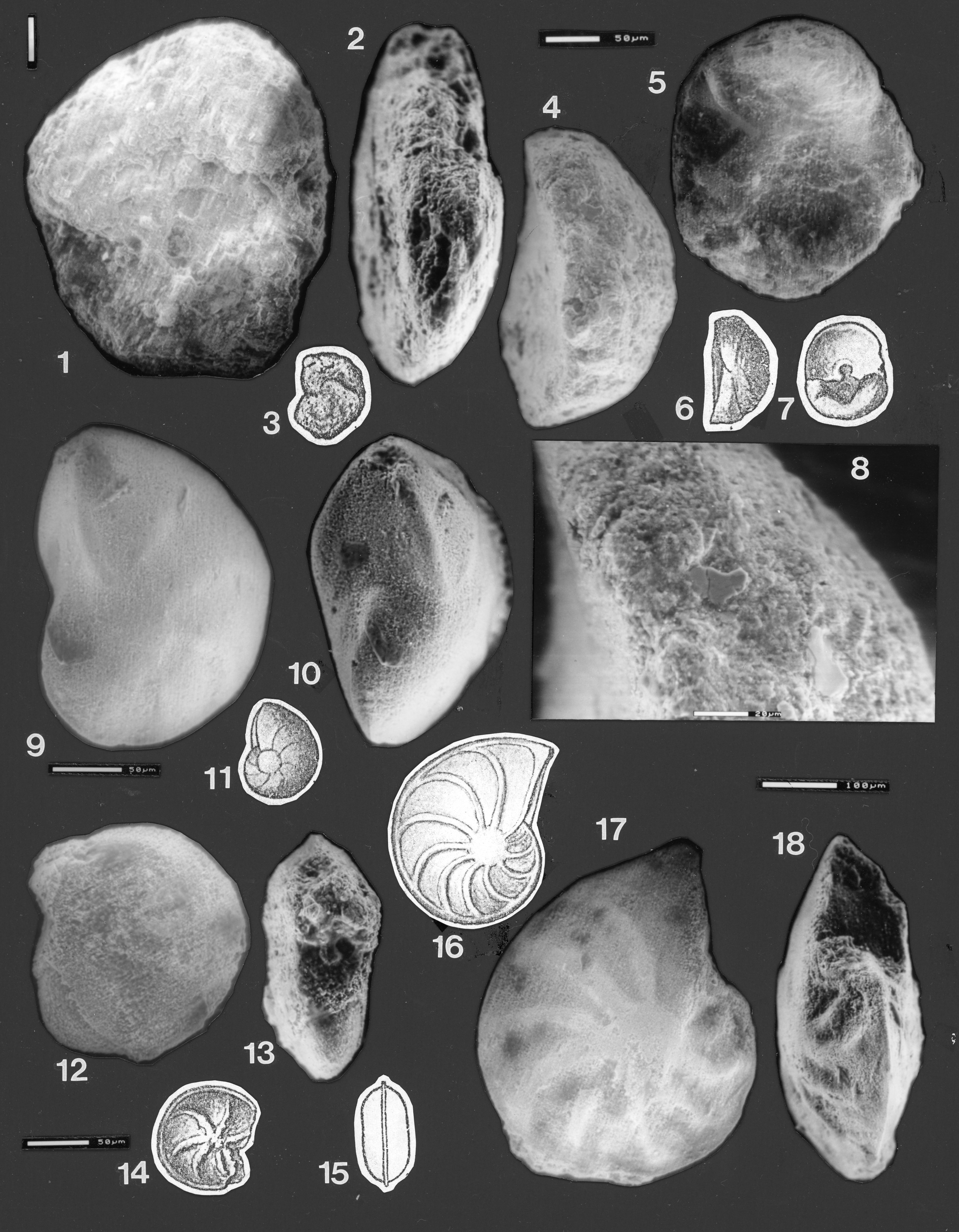
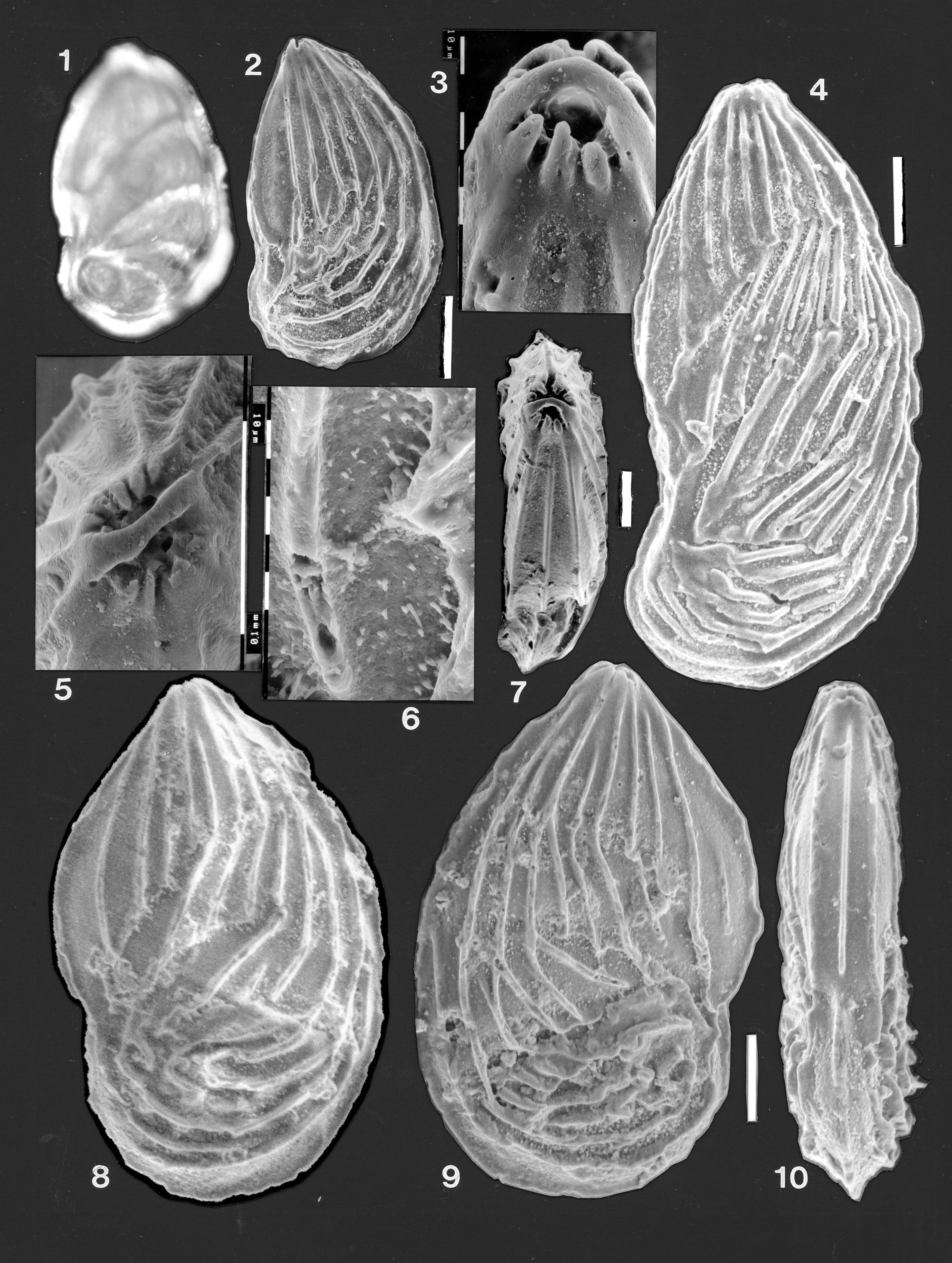
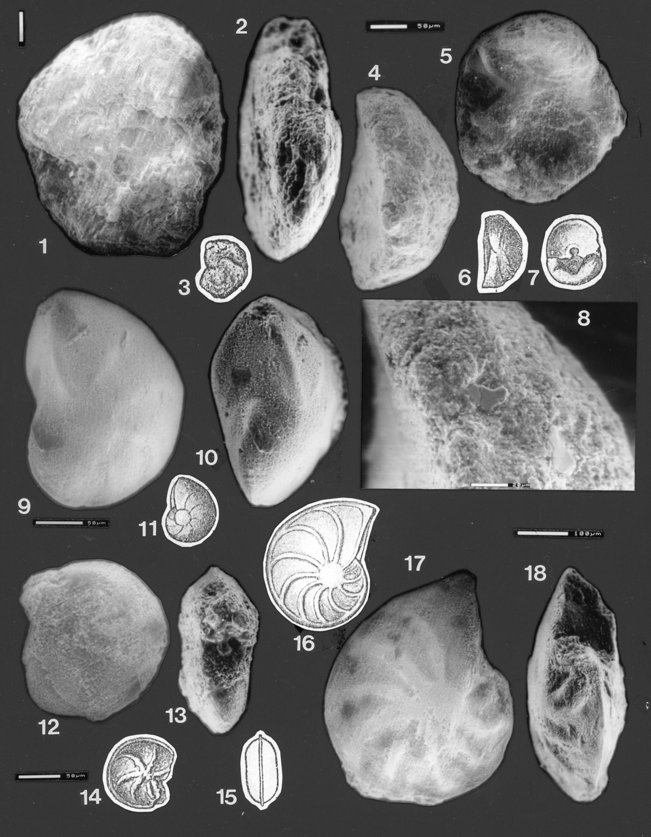
Fig. 2.1–3
1894 Haplophragmium neocomianum Chapman, p. 695, pl. 34, figs. 2a, b.
1904 Haplophragmium neocomianum Chapman, p.186–187, pl. 22, fig. 1.
Material examined
Chapman’s single specimen, Museum of Victoria specimen no. P2121.
Chapman’s description
From Chapman (1904, p. 186, 187): “This is a compressed, spiral, arenaceous form, having an irregular, sub-circular outline. The whorls of the test are involute, and the chambers are obscurely seen on the surface; but the latter are made apparent by moistening the test…”
Amended Description
Maximum height 0.41 mm; width 0.36 mm; thickness ~0.09 mm. A poorly preserved, planispirally coiled agglutinated test, composed of sand grains up to 40 microns across set in a finer orange-brown matrix. Outline sub-circular, slightly lobate, with about five poorly defined chambers in the final whorl. The umbilicus is depressed on one side (see Fig. 2.1), somewhat filled with rock matrix on the other. In edge view, compressed, somewhat broken, margin possibly rounded in the earlier part of the final whorl, narrowly rounded in the final chamber. The final chamber is damaged, with two elongated holes near the apex and on the final face, and a crack running along the top of the chamber (see Fig. 2.2). Chapman described the aperture of H. neocomianum as an arched slit at the base of the terminal chamber (fide Ellis and Messina 1940 et seq.). The ESEM photos strongly suggest that the holes visible in the final chamber are merely damaged wall, not an apertural opening. The aperture is partly obscured by glue and matrix but appears to be an elongated slit running along the basal suture of the final chamber. For this reason, the specimen is referred to Haplophragmoides Cushman 1910, rather than to Haplophragmium Reuss 1860.
Incertae Sedis
? Trochammina sp. (? mould)
Fig. 2.4–8
1904 Textularia crater Chapman, p. 187, pl. 22, figs. 2 and 2a.
Material examined
Chapman’s single specimen, Museum of Victoria specimen P2122.
Chapman’s description
From Chapman (1904, p. 187): “Test very short, laterally slightly compressed, and, therefore, sub-elliptical in cross-section. Aboral end rounded. Oral surface depressed, the apertural margin limbate. The margin of the last and penultimate chambers somewhat square with the upper surface of the test. Chambers few, obscurely seen on the surface of the shell, but the textularian arrangement may be made out without much difficulty. Texture finely arenaceous. Length of test, 0.14 mm. Greatest width, 0.3 mm… One specimen.”
Amended description
Length 0.27 mm; width 0.235 mm; height 0.125 mm. Outline round to slightly oval; concavo-convex in edge view. Surface roughened, reddish. Umbilical area depressed, initially partly filled with matrix. Both initially and after some cleaning of this area, no indication of an aperture (as shown in Chapman’s drawing) was seen. There is no indication of sutures or subdivision into chambers, both on the “spiral” side and on the “umbilical” side, except for a more lobate area (Fig. 2.5) at the top that resembles a final chamber. Chapman’s description of a “textularian” chamber arrangement was not seen, although there are some grooves on the surface, particularly in lateral view (Fig. 2.4) that obviously influenced his interpretation. There is no indication of cemented agglutinated wall structure in the light microscope or ESEM, the vari-coloured reddish surface resembling an internal mould (Fig. 2.4, 5, 8). This author believes that the specimen might be an internal mould of either a Trochammina or an aragonitic form, but also concurs with S. Revet’s opinion that, even after cleaning, it is not possible to distinguish chambers or to be sure that this is a foraminifera.
Suborder Lagenina Delage and Hérouard 1896
Family Vaginulinidae Reuss 1860
Subfamily Lenticulininae Chapman, Parr and Collins 1934
Genus Lenticulina Lamarck 1804
Lenticulina sp. cf. L. notus (Zaspelova 1948)
Fig. 2.9–11
not 1804 Lenticulites rotulata Lamarck, p. 188; (1806, pl. 62 (14), fig. 11); (fide Ellis and Messina 1940 et seq.)
1904 Cristellaria rotulata Lamarck; Chapman, p. 192, pl. 22, fig. 9.
not 1939 Cristellaria wisniowskii Myatliuk, p. 57, pl. 4, fig. 43.
1948 Cristellaria notus Zaspelova, p. 206, pl. 2, fig. 6a–b.
1955 Lenticulina wisniowskii (Myatliuk); Tappan, p. 54, pl. 16, figs. 2–3 only.
? 1985 Lenticulina tympana Grigelis, p. 73, pl. 1, figs. 11a–b, pl. 15, fig. 3.
? 2001 Subhercynella muensteri (Roemer 1839); Bartenstein & Malz, p. 35, pl. 3, figs. 53–56.
Material examined
Chapman’s single specimen, Museum of Victoria specimen P2132; Senckenberg XXVII 607a 30–35 of Bartenstein and Brand 1937.
Chapman’s description
From Chapman (1904, p. 192): “This widely distributed and persistent type is a well-known form in jurassic deposits, but it is not nearly as common as the more ornate type with secondarily thickened sutures…”
Amended description
Maximum diameter ~0.21 mm; maximum thickness near umbo ~0.1 mm. An involute coiled Lenticulina of about one and a half whorls; about 6.5 to 7 chambers in the final whorl. Outline not completely circular but slightly elongated towards the aperture. Maximum thickness at the low but distinct umbilical boss, but with the final chamber fairly wide and slightly inflated. Terminal face of the last chamber triangular, slightly concave, outlined by a low ridge. Sutures curved backwards, slightly but distinctly depressed, emphasised by a slight inflation of the chamber posterior to the suture. Aperture at the terminal angle of the final chamber, slightly produced but broken. Test opaque, earliest chambers not visible.
Remarks
Smooth Jurassic Lenticulina have few distinctive characteristics, and authors have followed two approaches, either referring such specimens to Lenticulina muensteri (Roemer 1839) or alternatively naming many new species that differ only subtly in test shape (eg. Cristellaria vulgaris Schwager 1867 (in Waagen 1867); Cristellaria notus Zaspelova 1948). Lenticulina muensteri, as examined in some European collections (eg. Senckenberg XXVII 607a 30–35), has more chambers in the final whorl (9 to 12, sometimes more) in addition to being considerably larger (0.5–1mm diameter). Chapman referred his specimen to the Cretaceous species Lenticulites rotulata Lamarck, but the latter has more chambers in the final whorl, and the drawing of that species (fide Ellis and Messina 1940) suggests that the coiling is more involute and that the chambers are less elongated towards the aperture than Chapman’s specimen. Cristellaria wisniowskii Myatliuk is slightly more flattened, particularly in the final chamber, and the sutures are described as flush. Chapman’s specimen has a wider final chamber, and the sutures are shallowly depressed. Lenticulina tympana Grigelis is similar in shape and number of chambers in the final whorl, but the sutures are flush and appear to be radial. Subhercynella muensteri (Roemer) of Bartenstein and Malz from the Bringo Cutting locality, Western Australia, are similar but are figured from broken specimens which do not show the slight depressions along the sutures, nor the low but distinct umbonal plug. The specimen is tentatively referred to the Upper Jurassic to Lower Cretaceous species Lenticulina notus (Zaspelova) on the basis of the shape, the umbonal disc, and the presence of seven chambers in the final whorl, but further study of this and similar small, smooth forms are needed for a satisfactory identification.
Lenticulina subalata (Reuss 1854)
Fig. 2.16–18
1854 Cristellaria subalata Reuss, p. 68, pl. 25, fig. 13a–b (fide Ellis and Messina 1940 et seq.)
not 1863 Cristellaria subalata Reuss, p. 76, pl. 8, fig. 10; pl. 9, fig. 1 (fide Ellis and Messina 1940 et seq.)
1904 Cristellaria subalata Reuss; Chapman, p. 193, pl. 22, fig. 10.
2001 Lenticulina subalata (Reuss); Bartenstein and Malz, p. 35–36, pl. 3, fig. 57.
Material examined
Chapman’s single specimen, Museum of Victoria specimen P2133.
Chapman’s description
No description but the following remarks were made by Chapman (1904, p. 193): “This species is also known from older tertiary strata, but is more commonly met with in the jurassic and lower cretaceous series…”
Description
Maximum diameter 0.45 mm; thickness at umbo ~0.18 mm. Moderately large, outline nearly circular, biconvex, involute Lenticulina, with about 9.5 chambers, gradually expanding in size, in the final whorl. Earlier chambers not visible. In side view, central raised umbo and prominent thick raised sutures, moderately curved. Aperture slightly produced, at the ventral angle. In edge view, fairly compressed, biconvex, umbonate. Periphery acute, with a thin keel, better developed on the earlier chambers in the last whorl. Terminal face broken, apertural details obscure.
Lenticulina sp.
Fig. 2.12–15
not 1855 Cristellaria prominula Reuss, p. 271, pl. 9, figs. 3a–b (fide Ellis and Messina 1940 et seq.)
1904 Cristellaria prominula Reuss; Chapman, p. 193, pl. 22, figs. 11, 11a.
Material examined
Chapman’s single specimen, Museum of Victoria specimen P2134.
Chapman’s description
From Chapman (1904, p. 193): “The distinctive features of this form are the presence of the sutural lines which mark the junction of chambers, less strongly curved on the septal face than in the nearly allied C. subalata; and the greater height of the chambers when compared with the species just mentioned”.
Amended description
Maximum diameter 0.18 mm, thickness 0.07 mm. This extremely poorly preserved specimen is heavily etched and has the wall entirely broken away over part of the whorl, revealing a yellow internal mould. Outline almost circular; test somewhat compressed, sides flat, umbilical area slightly depressed. Chambers arranged in an involute coil, earliest chambers not visible; final whorl consisting of about 7 chambers, slightly inflated, expanding very slowly in size. Sutures only slightly raised, thickened when seen in light microscope; final suture slightly depressed. The irregular outline of the sutures shown in Chapman’s drawing is due to small patches of calcareous matrix cemented onto the specimen, plus an irregular, “sugary” etching of the white sutures. In the ESEM photograph of the opposite side of the specimen, which has less matrix adhering, the highly etched and recrystallised nature of the wall is apparent (Fig. 2.12). The raised appearance of the sutures is partly due to the greater amount of etching of the chamber walls between the sutures, the sutures themselves being very low or even depressed. The periphery is narrowly rounded and defined by a blunt, very poorly preserved keel in the later chambers (Fig. 2.13). The aperture is broken and obscured by adhering fragments.
Remarks
This specimen is not considered to be conspecific with Lenticulina prominula (Reuss 1855) as it is relatively thicker, less lobate and has a more rounded margin. The extremely poor preservation does not permit any meaningful identification
Lenticulina quenstedti (Gümbel 1862)
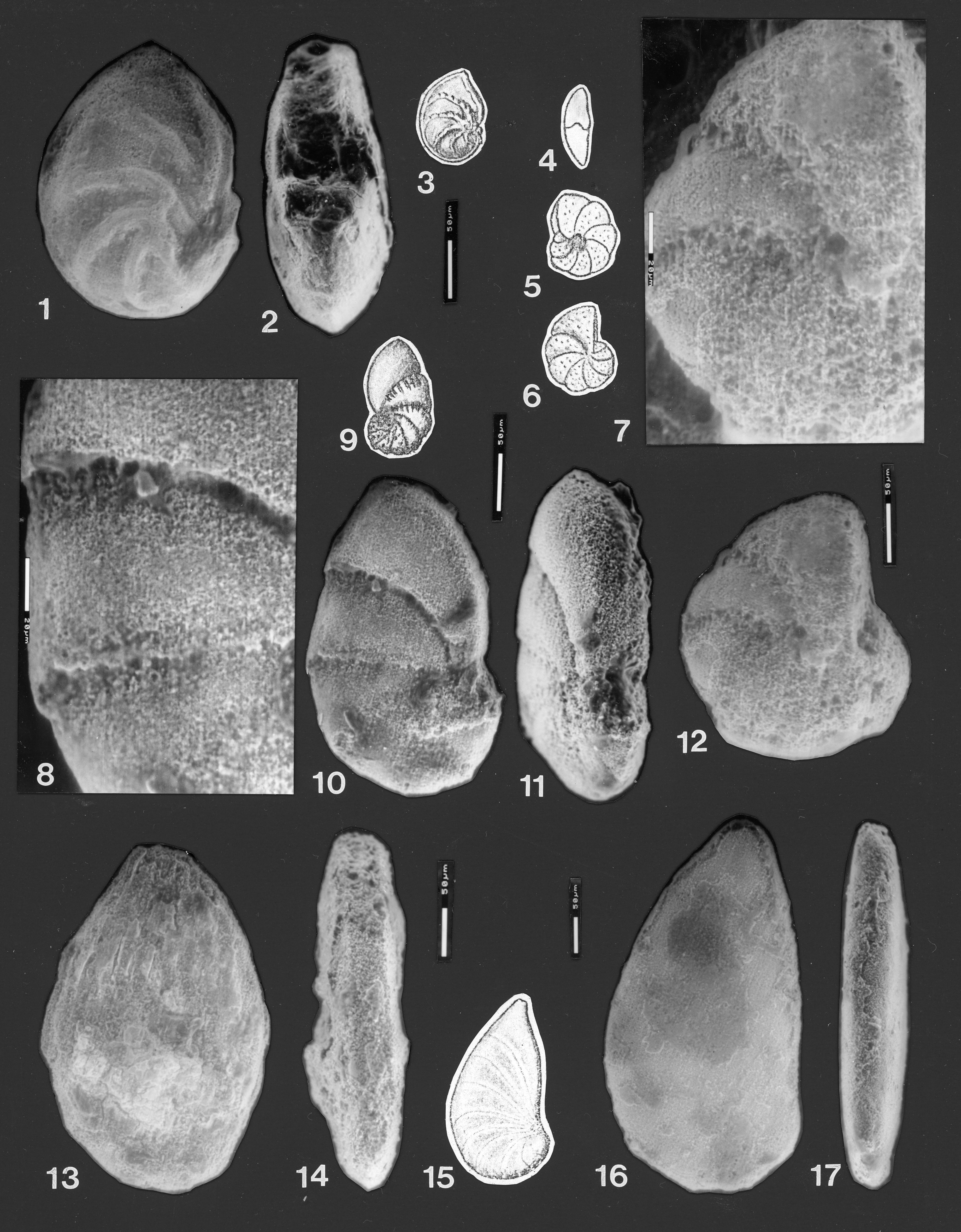
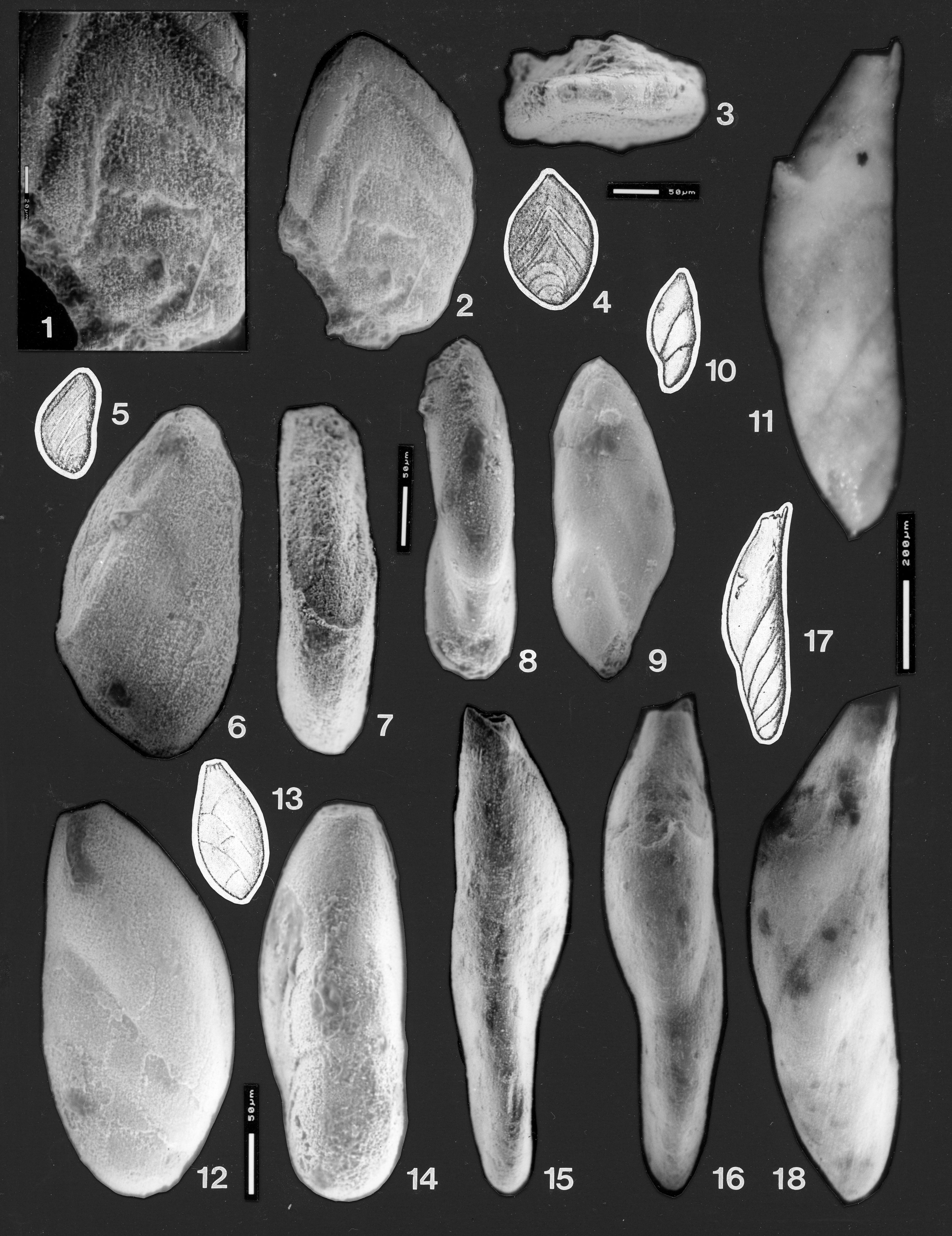
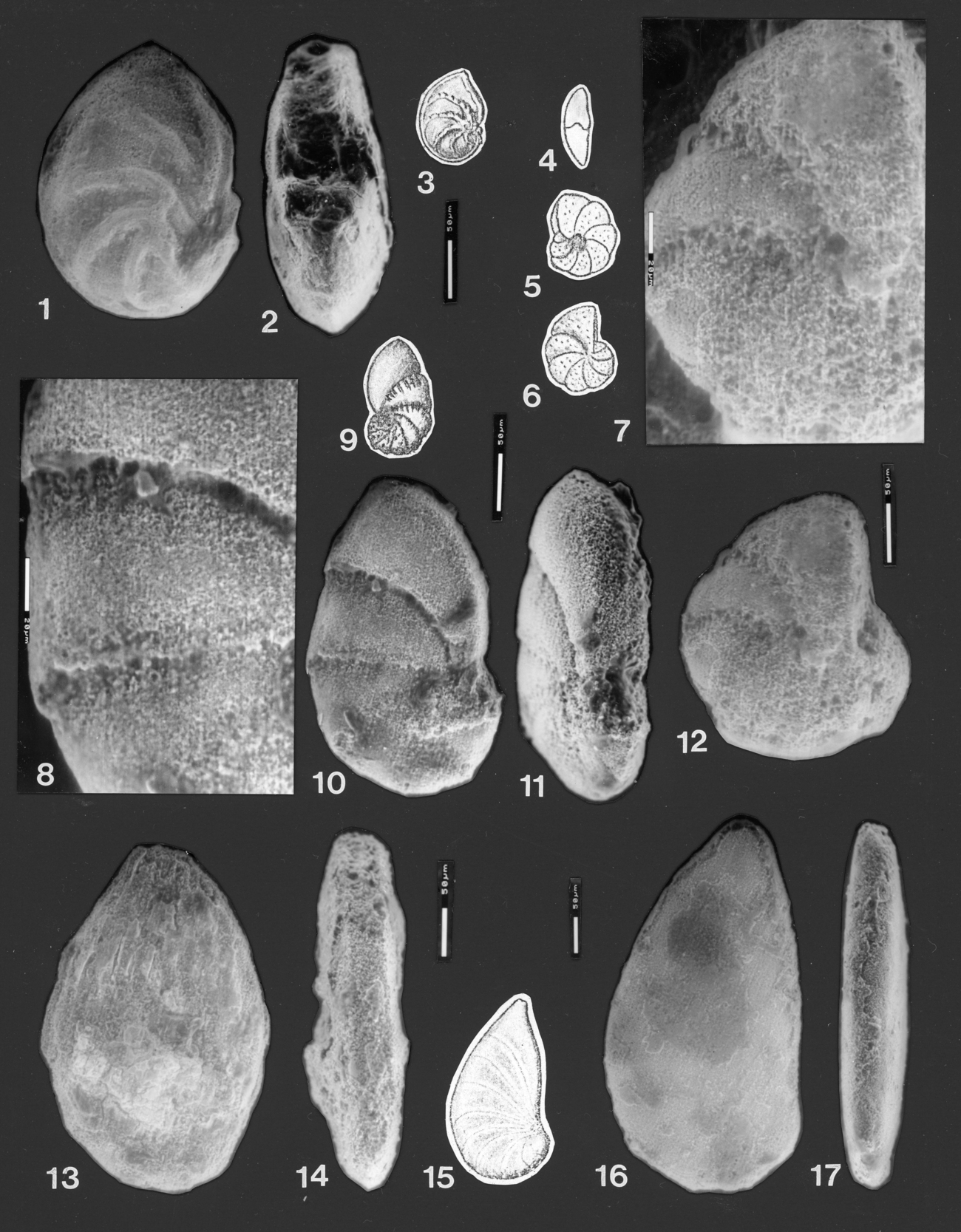
Fig. 3.1–3
1862 Cristellaria quenstedti Gümbel, p. 226, pl. 4, fig. 2a–b.
1890 ?Cristellaria decipiens Wisniowski, p. 222, pl. 10, figs. 5a–b, 11a–b (fide Ellis and Messina 1940 et seq.)
1904 Cristellaria decipiens Wisniowski; Chapman, p. 193, pl. 22, fig. 12.
1955 Lenticulina (Lenticulina) quenstedti (Gümbel 1862); Seibold and Seibold, p. 105, text fig. 3f–g, pl. 13, fig. 3.
2000 Lenticulina (Lenticulina) quenstedti (Gümbel 1862); Bartenstein, p. 407–409, pl. 1, fig 2a; pl. 4, figs. 39a–b.
not 2001 Astacolus volubilis (Dain 1958); Bartenstein and Malz, p. 47, text-fig. 9, fig. 12.
Material examined
Chapman’s single specimen, Museum of Victoria specimen P2135.
Chapman’s description
From Chapman (1904, p. 193, 194; for his C. decipiens): “This ornate cristellarian belongs to the group of which C. cassis, Fichtel and Moll. sp., forms a central type. The original specimens figured by Wisniowski, came from the upper jurassic (Am. ornatus beds) of Poland.”
Amended description
Maximum diameter 0.25 mm; thickness at last chamber 0.09 mm. Test oval, a planispiral coil of >6 chambers; test broken along the ventral margin, removing the last chamber and obscuring details of the earliest chambers (Fig. 3.1–2). Earliest chamber not clearly visible, possibly at the ventral margin, or possibly slightly internal from the margin. The only visible whorl consists of 6 chambers, with a seventh broken off; chambers triangular, increasing in width; the last chambers reaching back and touching the earlier ones. Chambers separated by curved, thickened, strongly raised sutures which die out towards the outer part of the chambers, but which are connected in the umbonal area by an irregular ridge that might have formed part of the perimeter of a circle, before the area was broken. The sutures do not join above the umbo in the manner shown in Chapman’s drawing (reproduced here as Fig. 3.3). Test shape moderately compressed. Peripheral margin somewhat eroded but generally acute, on some chambers with a poorly developed marginal keel. Aperture and entire ventral face broken off. The “ornament” shown on the last two sutures in Chapman’s drawing was small sugary lumps of matrix, some of which detached from the specimen when it was mounted for ESEM photography; they are not costae like those seen in Lenticulina volubilis Dain 1958 (in Bykova et al. 1958). The opposite side from that photographed has additional matrix adhering to it.
Remarks
The species Lenticulina decipiens (Wisniowski), used here by Chapman, and Lenticulina polonica (Wisniowski) appear to be indistinguishable. Wisniowski separated his species Cristellaria decipiens from his species C. polonica by the absence of the “distinct semiannular ridge around the umbo which usually occurs in the species C. polonica, although there are exceptions to this rule in that species…” (fide Ellis and Messina 1940 et seq.) Bartenstein (2000), in a detailed study of Lenticulina polonica and Lenticulina quenstedti (Gümbel 1862), concluded that L. polonica was a synonym of L. quenstedti, a view that this author agrees with.
Seibold and Seibold (1955) designated a neotype for Lenticulina quenstedti (Gümbel 1862) and their drawing of this specimen suggested that the umbo was surrounded by a nearly continuous raised ring that joins the inner ends of the raised sutures. Bartenstein (2000) refigured the neotype by SEM and showed that, on the opposite (right) side of the test, the raised ring around the umbo is not continuous, joining only the last four sutures, with a break, then a short ridge linked to one and possibly two of the earlier sutures. This discontinuous ring around the umbo is very similar to the partial ring seen in Chapman’s specimen, which links four sutures. The identification is complicated by damage to Chapman’s specimen in this area (there is a hole in the test wall), and by the very poorly preserved opposite side of Chapman’s specimen. The variability of the ringlike ornament around the umbo in L. quenstedti is mentioned by many authors. Australian specimens of L. quenstedti are not common, but those specimens found also display great variability in the continuity of the ring-like ornament.
?Lenticulina minuta (Bornemann 1854)
Fig. 3.8–11
?1854 Cristellaria minuta Bornemann, p. 42, pl. 4, figs. 37a–b.
1904 Cristellaria daintreei Chapman, p. 194, pl. 22, fig. 13.
not 1955 Astacolus daintreei (Chapman); Tappan, p. 55, pl. 18, figs. 13a–b.
Material examined
Chapman’s single specimen, Museum of Victoria specimen P2136.
Chapman’s description
From Chapman (1904, p. 194, for C. daintreei sp. nov.): “Specific characters. - Test ovate-elongate, compressed, nine chambers visible, those forming the spiral portion sparsely granulate to tuberculate. The sutural depressions of the last three chambers bridged over by about seven short longitudinal costae. The last chamber is somewhat inflated. Length 0.46 mm; greatest width 0.2 mm. Affinities. - The nearest analogue to this species is C. gemmata, Brady. Both forms have a compressed test, but the ornamentation of C. daintreei differs essentially in the semi-costate surface marking of the last few chambers. In the character of the ornament C. daintreei also resembles some extreme forms of C. fragraria, Gümbel sp. (= C. wetherelli, Jones sp.) in which the test is strongly compressed, and the later chambers costate.”
Amended description
Test elongated, ovoid, compressed. Coiled in a single planispiral whorl, consisting of about nine chambers; initial chamber obscured by adhering matrix, which Chapman described as “granulate to tuberculate” test. Chambers 2–6 close-coiled, expanding slowly in size; chambers 7 to 9 expanding rapidly in height, and moderately rapidly at the dorsal margin so that the test begins to flare (Fig. 3.10); however, the last chamber is still in contact with the initial whorl. The final chamber is slightly more inflated than the earlier chambers. The peripheral view is compressed, with narrowly rounded periphery and almost flat sides (Fig. 3. 11). Sutures between the early chambers thickened, flush; but between the last two chambers, deeply depressed, partly because the test wall of the later chambers is largely dissolved. The last two chambers appear to be an internal mould. There are no sutural bridges or costae across the sutures of the last three chambers (Fig. 3.8, 10). These features, referred to by Chapman as diagnostic for the species, in the ESEM can be seen to be heavily etched remnants of sutural material (see Fig. 3.8). There are no costae or genuine sutural bridges on either side of the single very poorly preserved specimen, which was examined on both sides.
Remarks
The holotype (and only specimen) of Cristellaria daintreei Chapman does not exhibit the “semi-costate surface markings” nor the bridges of costae across the sutures that were described, nor the “granulate to tuberculate” initial portion. It appears to be a heavily etched, infilled specimen that is now partly an internal mould, and has adhering matrix on the earliest chambers. The part of the wall that remains is smooth. It is therefore considered that the species name daintreei should be relinquished in favour of a tentative identification as Lenticulina minuta (Bornemann).
Lenticulina sp.
Fig. 3.4–7, 12
not Anomalina wüllerstorfi Schwager, 1866, p. 258, pl. 7, figs. 105, 107 (fide Ellis and Messina 1940 et seq.)
1904 Truncatulina wuellerstorfi Schwager, 1866; Chapman, p. 198, pl. 22, figs. 24, 24a, 24b.
Material examined
Chapman’s single specimen, Museum of Victoria specimen P2148.
Chapman’s remarks
From Chapman (1904, p. 198, 199, for his T. wuellerstorfi): “…The example before us shows all the characters of the species, and especially the tendency of the chambers to overlay one another at the sutural lines, whereas in T. lobatula the separate chambers are more inflated.”
Description
Maximum diameter 0.21 mm. Outline more or less circular, sides flattened, margin narrowly rounded. The last whorl consists of a planispiral coil of about 7 chambers. Sutures curved, depressed. Aperture not seen. Fragments of matrix occur on the specimen in the umbonal area. The test wall is severely etched and totally dissolved away in places.
Remarks
The asymmetry of coiling shown by Chapman was not evident during this examination. This appears to be a very badly preserved Lenticulina.
Genus Spincterules Loeblich and Tappan 1987
Type species. Spincterules anaglyptus Loeblich and Tappan 1987, nomen novum.
Spincterules seminudus (Chapman 1904)
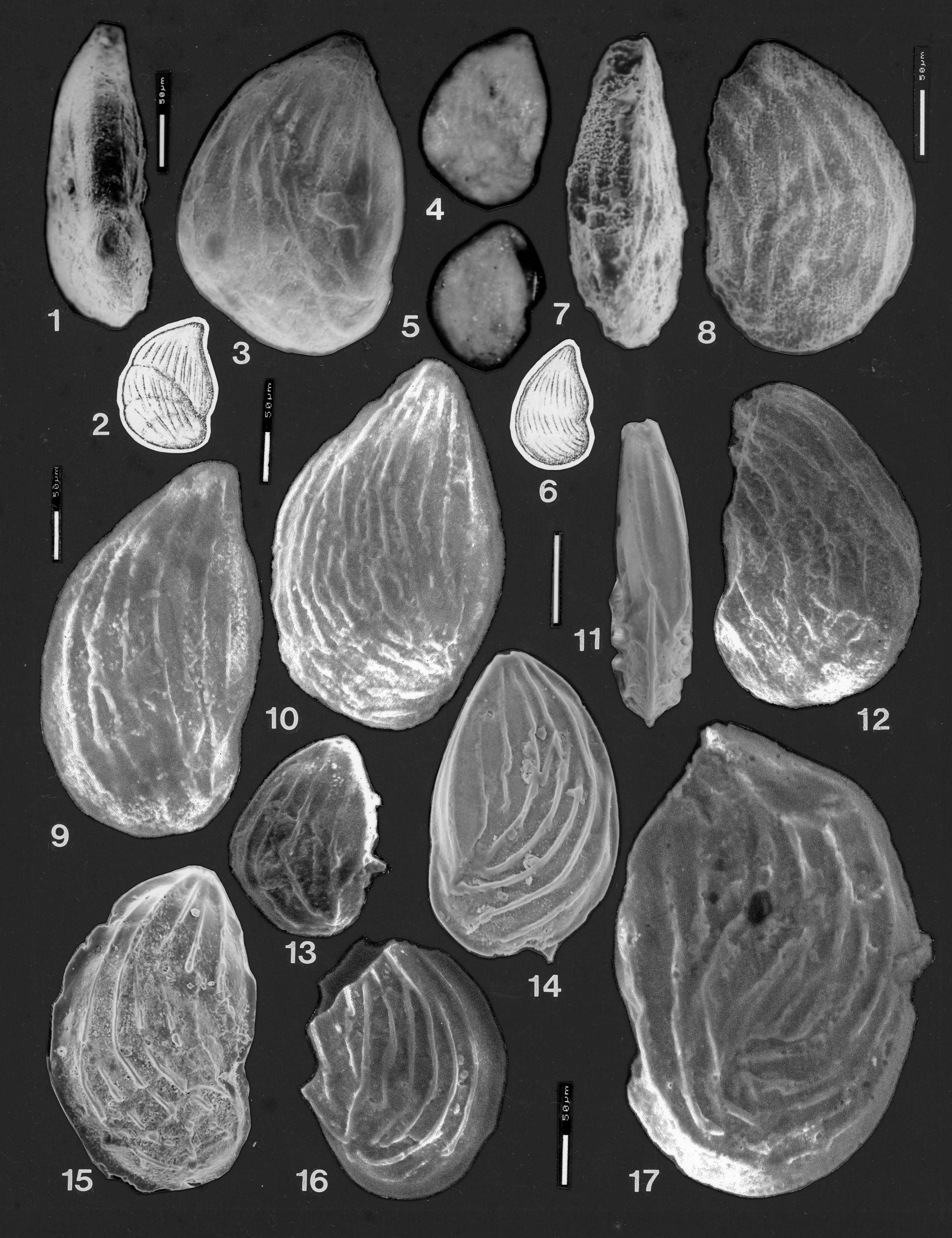
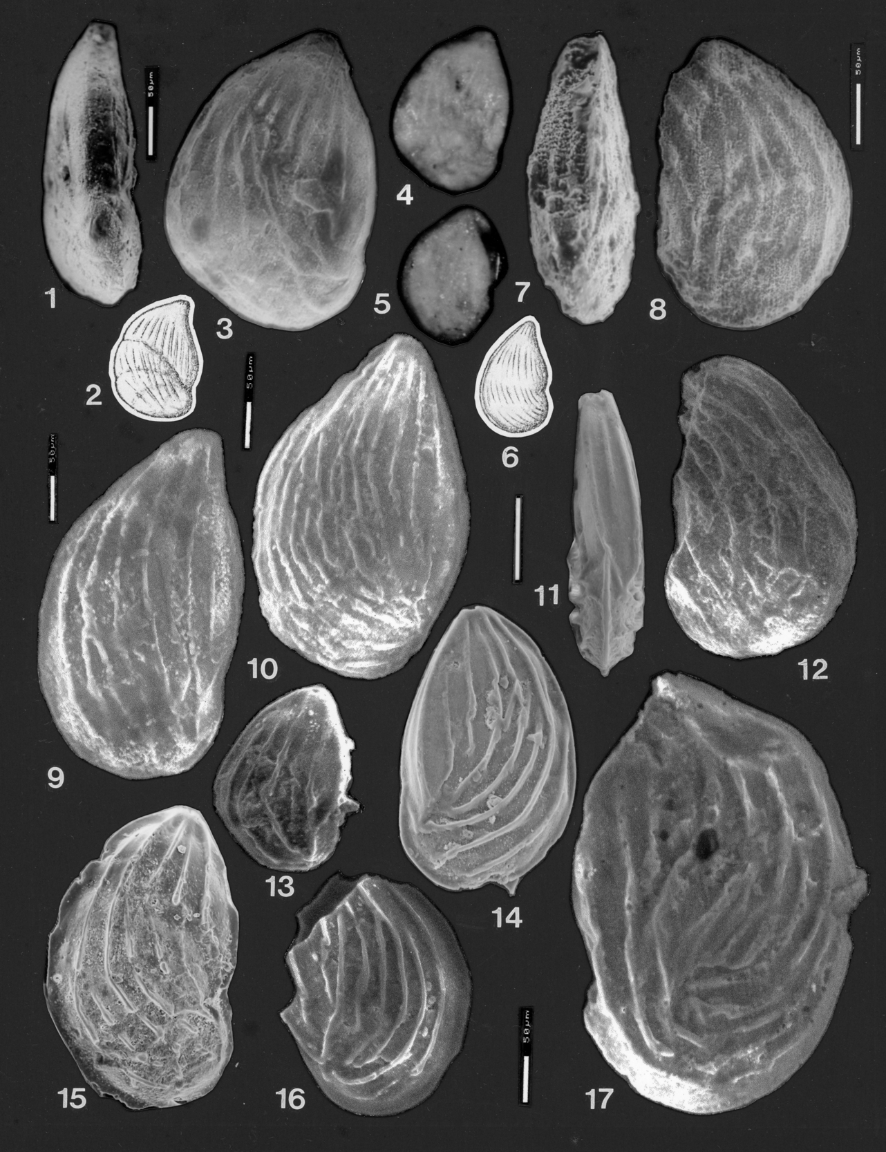
Fig. 4.1–17; Fig. 5.1–10
not 1798 Nautilus costatus Fichtel and Moll, p. 47, pl. iv, figs. g, h and I (fide Ellis and Messina 1940 et seq.)
not 1842 Planularia compressa von Hagenow, p. 569.
not 1846 Cristellaria compressa d’Orbigny, p. 86, plate 3, figs. 32, 33.
not 1883 Robulina semi-nuda Terquem, p. 366, pl. 41, fig. 13a–b (fide Ellis and Messina, 1940 et seq.)
1904 Cristellaria costata, Fichtel and Moll sp., var. compressa, nov. Chapman, pp. 194–195, pl. 22, fig. 14, Museum of Victoria Specimen P2137.
1904 Cristellaria costata, Fichtel and Moll sp., var. seminuda, nov. Chapman, p. 195, pl. 22, fig. 15, Museum of Victoria Specimen P2138.
1963 ?Lenticulina collignoni Epistalié and Sigal (part), pl. 9, figs. 4a–4d only.
not 1994 Spincterules compressus Loeblich and Tappan, pp. 70–71, pl. 126, figs. 8– 13; pl. 134, figs. 8–9.
1995 Lenticulina (Vaginulinopsis) reticulosa (ten Dam, 1946); Bartenstein, p. 11, pl. 3, fig. 36 only.
2001 Lenticulina (L.) gerzenensis geraldtonensis Bartenstein and Malz, p. 40, pl. 4, figs. 82–87.
2001 Roemerelina incohata Bartenstein and Malz, p. 42, pl. 4, figs. 90–91.
2001 ?Roemerelina incohata Bartenstein and Malz, p. 42, pl. 4, fig. 89.
Holotype
Museum of Victoria P2138.
Comparative material examined
The holotypes of Chapman’s species Cristellaria costata var. compressa, (Museum of Victoria specimen P2137) and Cristellaria costata var. seminuda, (Museum of Victoria specimen P2138) have been examined and are figured herein. Numerous specimens from the Bringo Cutting, type locality of Lenticulina (L.) gerzenensis geraldtonensis Bartenstein and Malz, figured herein.
Chapman’s descriptions
From Chapman (1904, p. 194, 195 for specimen P2137): “Cristellaria costata, Fichtel and Moll sp., var. compressa, nov.” “The present variety, compressa, is a strongly compressed form. The spiral commencement is not well developed, and in this, as in other features, such as the broad curvature of the septa, it closely approaches the vaginuline style of test. The longitudinal costae are fairly well developed but are somewhat interrupted near the sutural depressions. Length of specimen 0.4 mm; greatest width 0.3 mm.”
From Chapman (1904, p. 195 for specimen P2138): “Cristellaria costata, F. and M. sp., var. seminuda, nov.”, holotype of Spincterules seminudus (Chapman). “This variety differs from the type [ie. Cristellaria costata Fichtel and Moll] in the interruption of the longitudinal costae on the central area of each chamber. The test is also more elongate, with fewer chambers, and is less inrolled than the type-form. The peripheral margin is bordered by a narrow keel. Length of test, 0.38 mm; width, 0.22 mm.”
Amended description of P2137
Length ~0.3 mm; greatest width ~0.22 mm; thickness ~0.07 mm. Specimen tear-shaped in outline, proloculus at periphery, and the following four chambers in an evolute partial coil (Fig. 4.3–4). Chambers two and three are eroded, with much of the chamber wall missing. The final (sixth) chamber reaches back to touch the proloculus. Aperture produced but severely damaged. A poorly defined forward-facing part of the aperture appears to be largely filled with debris (Fig. 4.1). Test highly compressed with flattened sides; periphery somewhat angular on dorsal margin, eroded in part, with remnants of a small peripheral keel in places. “Ventral” face of the final chamber is outlined by two ridges, now almost completely eroded, one ridge marked by a groove (Fig. 4.1). The septa are curved, poorly visible, and almost flush, except for the last, which is depressed. The ornament consists of a poorly preserved peripheral keel and on each side of it, two costae which run continuously (apart from damaged areas) around the margin of the lateral faces of the chambers (Fig. 4.3). There are seven costae on each lateral face of the final chamber. The costae are straight, radiating from the apertural area back to the previous suture. Here they are interrupted in the slightly depressed final suture. Several costae on the ventral part of the final chamber do not extend back onto the previous chamber. The more dorsal costae continue onto the other side of the suture on the previous (fifth) chamber, where they intersect a second set of costae at an angle of 30˚, which radiate from the position of the aperture of this chamber. This set of costae continue back onto the fourth chamber, across an almost flush suture. The costae on the third and second chambers are missing, due to damage causing removal of the wall, and Chapman’s drawing of the costae is inaccurate on this point. One costa continues onto the proloculus.
Remarks
This abraded specimen, due to its poor preservation, does not fully reflect the typical adult morphology or ornament.
Description of P2138
This is the holotype of Spincterules seminudus (Chapman). Maximum diameter 0.26 mm, width 0.18 mm, thickness ~0.05 mm. Test outline ovoid, highly compressed, keeled, lateral sides almost flat, ornamented with prominent costae. Chambers in an evolute coil of seven or eight chambers forming a single whorl; earliest chambers cannot be discerned due to poor preservation (Fig. 4.5, 8). The last preserved chamber is broken off near its basal suture and is partly filled with matrix but is beginning to uncoil. The base of this chamber is only questionably in contact with the initial chamber. Sutures slightly depressed, but with a slightly raised area of chamber anterior to the suture on the last three chambers, over which the costae thicken, to form indistinct radial ridges (Fig. 4.8). Costae longitudinal, radiating from the location of successive apertures, back to the previous suture; sometimes joining previous costae, and sometimes crossing them at acute angles. A continuous curved costa outlines the edge of the lateral face, parallel to the dorsal margin. The periphery is subacute and damaged on the earlier part of the test, but has a well-developed keel preserved on the last 3.5 chambers. There is also a faint suggestion of a keel ridge at the base of the apertural face on the damaged last chamber (Fig. 4.7). Aperture broken away; a circular hole is present at the dorsal angle of the last chamber, beneath where the aperture would be located.
Remarks
This is a rather poorly preserved, broken specimen, with the longitudinal costae worn down in places to the level of the chamber surface, producing a discontinuous series of radial ridges in front of the sutures. This specimen has thicker costae and a more prominent dorsal keel than C. costata var. compressa. Chapman’s drawing of seminuda (reproduced as Fig. 4.6) is inaccurate. The initial coil is not differentiated from the outline of the later chambers, the later chambers are broken away leaving a ragged edge, and the longitudinal costae are not interrupted as shown, but are fairly continuous, although considerably abraded or weathered, particularly on the earliest chambers, on which only two costae are poorly discernible.
Remarks on taxonomic nomenclature of P2137 and P2138
Genus and species names: Chapman assigned both his specimens to Cristellaria costata (Fichtel and Moll), as new sub-species. The name Nautilus costatus Fichtel and Moll 1798, is a junior homonym of Nautilus (Orthoceras) costatus Batsch 1791, according to Loeblich and Tappan (1987), who renamed the Fichtel and Moll form Spincterules anaglyptus Loeblich and Tappan 1987, nomen novum.
Sub-species name: The sub-species compressa is raised to species rank because the present form is not closely related to the Recent species Cristellaria costata Fichtel and Moll (= Spincterules anaglyptus Loeblich and Tappan). Because the name Cristellaria compressa had already been used for other species within Cristellaria by several authors prior to Chapman, the name used by Chapman for this subspecies was a primary homonym and was therefore not available to him. It cannot therefore be used for Chapman’s species, even though that species is now considered to belong within the separate genus Spincterules (ICZN Article 57.2).
Chapman also described Cristellaria costata var. seminuda, (Museum of Victoria Specimen P2138), also from a single specimen. The species name seminuda is available for Chapman’s form because the name semi-nuda was ascribed to the genus Robulina d’Orbigny 1826 by Terquem in 1883. Transfer of Robulina to the genus Cristellaria (by Stache in 1864) makes Cristellaria seminuda Chapman 1904 a secondary homonym (Article 53.3) of Cristellaria (Robulina) semi-nuda Terquem 1883, but as the two are no longer considered to be congeneric, the junior species name seminuda of Chapman is not to be rejected (ICZN Article 59.2) but is available for the present species. This new taxon seminuda should, as for the species compressa Chapman, be placed within Spincterules (see Discussion).
Both “compressa” (Specimen 2137) and seminuda (Specimen 2138) are subjectively the same species, so that the correct name to apply to both specimens is Spincterules seminudus (Chapman, 1904). The specimens are within the range of variation of the abundant costate species of Spincterules that occurs in the Bringo Cutting locality (type locality for Lenticulina (L.) gerzenensis geraldtonensis Bartenstein and Malz). Both specimen size and degree of continuity of the costae are variable in this species (see Figs. 4 and 5).
Generic assignment: The peripheral profile is highly compressed and with flat sides, hence an assignment to Planularia would be used, were it not for the fact that the type species of Planularia is unornamented except for a single ridge along the dorsal margin. The present form is assigned to the genus Spincterules Loeblich and Tappan 1987 (within the subfamily Lenticulininae Chapman, Parr and Collins 1934) based on its costate ornament that radiates from each successive aperture; the compressed, non-umbonate test; the depressed sutures; and carinate periphery. The holotype of the Recent Spincterules compressus Loeblich and Tappan 1994, is morphologically almost identical to some specimens of Chapman’s Jurassic S. seminudus, except for the less slit-like apertural modification of the latter.
Discussion
Both Cristellaria costata var. compressa Chapman and Cristellaria costata var. seminuda Chapman fall within the range of variation seen in the large suite of S. seminudus present in the Bringo Cutting samples. The latter vary greatly in the details of their costae and in the architecture of the complex aperture (described below). If each variant of ornament in this material were to be described as a separate species, at least ten separate “species” would need to be established. When a large number of specimens are seen, it is obvious that small variations occur in every specimen, there being no two specimens that are identical (see below). Taken as a whole, S. seminudus with its prominent costae and heavy keel around the entire specimen (including usually the apertural face) is readily distinguished from other ornamented Lagenina in the same assemblage and does not intergrade with any other form.
Description of supplementary specimens
These specimens from the Bringo Cutting locality are conspecific with S. seminudus. (These specimens are housed in the West Australian Museum, Perth, under the catalogue numbers WAM 2026.42 to WAM 2026.54). Test large, up to 1 mm in length, commonly ~0.6 mm in length; oval outline, tapering towards the aperture; strongly compressed, with flat sides and chambers very slightly or not inflated.
Chamber arrangement initially loosely coiled, later uncoiling and becoming rectilinear. Evolute coil; all chambers are visible in many specimens. Proloculus large and oval, usually on the periphery or very near to it; possibly these specimens are the megalospheric form (Fig. 5.1, 2). Some larger specimens have a very small proloculus, one chamber removed from the periphery, and concealed in a slightly involute coil. This may be the microspheric form. Early chambers in an evolute or partial coil of one whorl or less, of five to eight triangular chambers. Later chambers in adult specimens uncoiling, with one to three chambers not in contact with the coiled part. Chambers increasing rapidly in size at the dorsal margin in the coiled portion in the first five to six chambers. In the uncoiled portion, there is only a slight increase in chamber height, so that the later uncoiled chambers slope back towards the initial coil, even if not reaching it. Proloculus variable in size (see above), and globular to ovoid in shape, often distinctly inflated. All subsequent chambers with flattened sides, not inflated. Sutures curved; slightly depressed to flush with the chambers in the early coiled portion of test; depressed in later portion.
In edge view, highly compressed and with flat sides. Periphery sub-acute, marked by a well-developed sharp keel extending the full length of the test, from the aperture, down the dorsal margin and extending to the earliest chamber; then extending upwards on the apertural face of the final chamber, terminating just below the aperture in a smooth area, below the radiate ridges of the aperture (Fig. 4.11; Fig. 5.7, 10).
Aperture terminal, situated asymmetrically towards the dorsal margin; produced slightly to prominently on a short tapering neck. The aperture itself is highly variable between specimens, but is basically a central circular opening, surrounded by radiate ridges (which are the ends of costae; see below). Some of the radiate ridges join in the centre to form bridges of various shapes across the aperture. There is no uniformity as to which of the ridges (if any) have fused in this way, so that bridges may be diagonal (Fig. 5.5) or perpendicular (Fig. 5.3, 7) to the final (ventral) margin of the chamber or may form a bridge with a bend in the middle. Not all apertures are bridged, some being entirely radiate. In damaged apertures (which are present in the great majority of specimens), the bridged elements are broken away, so that no estimate of the number of bridged versus radiate apertures could be made. The apertural characteristics of the species can best be described as produced, radiate, with the apertural opening variably subdivided by bridges formed by extensions of two of the costae. There is no uniform pattern as to which costae form these extensions. The aperture is not markedly extended down the frontal face of the chamber, although bridging produces an appearance of subdivision of the aperture into a frontal and an apical portion (as is also the case in some species of Lenticulina).
Wall structure calcareous, imperforate on margins, perforate on chamber sides between ornament of two types. Ornament very striking, prominent, consisting of two types of costae. The first are the continuous peripheral ridges, of which one forms a dorsal keel or flange, and two others form a continuous ridge close to the dorsal periphery on each side of the test, these ridges and the keel extending back to the proloculus (Fig. 4.16–17; Fig. 5.2, 4, 8, 9). The apertural face (or the usually truncate, flattened lateral face of the last one or two chambers on the “ventral” side), between the aperture and the initial chamber, is also outlined by two ridges, and has a central keel (not as high as the dorsal margin keel) running from below the aperture along the periphery down to the keel on the proloculus (Fig. 5.7, 10). In planispirally-coiled specimens this ridge joins the keel. In other specimens with twisted, imperfect planispiral coiling, the central ridge, and the ventral margin, are twisted to one side (Fig. 4: 11). The second type of ridge are the less continuous, narrow, high costae which occur on the flat chamber sides. In some specimens these costae can be seen to be internally hollow (Fig. 5.6). The ornament commences with several short costae with a random orientation on the proloculus (Fig. 4.9, 15; Fig. 5.2, 4, 8). By the third chamber the costae have become three or four in number, subparallel to the periphery and converging towards the aperture of each successive chamber (Fig. 4.14, 16; Fig. 5: 4). Costae on the flat sides of later chambers are semi-continuous with, or branch from, earlier costae, but change direction at each suture so that the costae always point in straight lines towards successive apertures (Fig. 4.12, 13, 14, 17; Fig. 5.2, 4, 8, 9). It is this series of radiating, straight costae that change direction, which gives the species a distinctive appearance. One to three additional costae are inserted at the sutures of later chambers; some of these die out before reaching the aperture. In coiled chambers there are approximately five costae on the flat chamber face, and in uncoiled chambers, six to eight costae (including 2–3 weak or very short ones) on the flat chamber face. Most of the costae, including the dorsal “keel” and the two lateral costae outlining the ventral face, continue up to the aperture and form the “radiate” ridges around the apertural opening (Fig. 5.7). The central ridge on the ventral face dies out before reaching the aperture (Fig. 4.11; Fig. 5.3, 7, 10). A further type of ornament also occurs on the chambers. Between the costae on the flat sides of the chambers, a spikey, pustulose ornament covers the chamber surface between the scattered pores (Fig. 5.6). This type of ornament is heaviest on the earliest chambers, and along the slightly depressed sutures, and is weakest on the youngest chamber, and virtually absent on the ventral periphery of the youngest chamber. The granular ornament is very poorly developed on juvenile specimens (Fig. 4.11, 14). For the above reasons, this spikey granular ornament is believed to be formed by secondary lamination during chamber addition, rather than being a diagenetic effect. However, in some poorly preserved specimens, there is an obvious diagenetic overgrowth on top of the granular ornament.
The pattern of costae varies between specimens. In the simplest pattern, where the specimen is only slightly coiled, five or six costae extend in a subparallel, semicontinuous vertical pattern from chamber number two, to the aperture (Fig. 4.14). Other specimens have a similar vertical pattern, but the costae show convergence towards earlier chamber apertures (Fig. 4.9). With an increasing degree of curvature of the initial coil, the earlier apertures tend to be less overlapped by subsequent chambers, and the costae can be seen to converge and join below the concealed aperture. Irregularities in the size increase of chambers also produce irregularities in the pattern of costae, with short ridges inserted in places, some costae joining or branching, and extensions of costae from the latest chambers running down to coalesce with the costae on the proloculus (Fig. 4.17). Some specimens (Fig. 4.10) have an increased number (up to 9) of finer costae per chamber. Some specimens (Fig. 4.15) have discontinuous costae that terminate completely at each suture, and this does not appear to be caused by post-mortem specimen damage. Occasional specimens have been seen where a specimen has been broken, then has regrown. Some specimens have a different pattern of costae, and different degree of coalescing of costae, on opposite sides of the same test (Fig. 5.8–9).
Comparison with other species
The ornament of S. seminudus differs from that of Planularia eugenii Terquem of Coleman (1982; specimen B.G.S. MPK 2409 = Coleman’s plate 2, figure 14), which is similar in shape. The latter specimen has continuous ornament across the sutures, rather than the discontinuous, oblique and aperturally radiating ornament of S. seminudus.
Marginulina omskiensis Balakhmatova 1960 (in Glazunova et al. 1960), from the Maastrichtian (uppermost Cretaceous), appears to be slightly more inflated on the lateral sides of the chambers. It has a variable costate ornament which parallels the periphery, but which is also seen to converge towards the aperture, as in S. seminudus. The “apertural face” (= ventral margin of the last chamber) has three or four narrow “keels”.
Spincterules seminudus is similar in chamber arrangement and test compression and ornament to the Recent species Spincterules compressus Loeblich and Tappan 1994. Both species are highly compressed, with costae which always radiate in nearly straight lines from the aperture of each successive chamber, thus changing angle sharply at most of the sutures. The differences between the species comprise: (1) There is generally less continuity of the costae on the flat chamber sides of S. seminudus, some of the costae ending at the depressed sutures while others join costae on the next chamber. In one paratype of S. compressus (USNM 470280: Loeblich & Tappan 1994, their Plate 134, figures 8, 9) the sutures are raised, a feature not seen in S. seminudus. (2) In S. seminudus the final chamber has a flattened ventral face outlined by two costae and ornamented by a single central costa (rarely two), usually running most of the length of the chamber and disappearing below the aperture. Spincterules compressus has a short costa on the early part of the final face in the photograph of the holotype (USNM 470286: Loeblich and Tappan 1994, their Plate 126, figures 12, 13) and is shown with a smooth final face in the drawing of a paratype (USNM 470287: op cit. their Plate 126, figure 9). It is described as having a smooth apertural face. In another paratype (USNM 470280: Loeblich and Tappan 1994, their Plate 134, figure 9) the costae from the flat chamber sides appear to curve around onto the final chamber face, which is thus not differentiated from the chamber sides. This paratype appears to represent a different species. (3) The dorsal keel on S. seminudus appears to be much higher than that illustrated for S. compressus, which is described as having a rounded periphery. (4) The aperture in S. compressus is described as radiate, with an elongated slit extending down the apertural face of the last chamber. In S. seminudus the aperture is highly variable between specimens, but is basically radiate, with some of the radial elements joining in the centre to form bridges (see Description). There is no elongated slit, although in a few bridged specimens, the triangular anterior part of the aperture extends a very short distance down the apertural face.
Subfamily Marginulininae Wedekind 1937
Genus Astacolus Montfort 1808
? Astacolus renominatus Subbotina, Datta and Srivastava 1960
Fig. 3.13–17
not 1846 Cristellaria lanceolata d’Orbigny, p. 89, pl. 3, figs. 41–42.
1865 Cristellaria lanceolata Schwager, p. 130, pl. 6, figs. 13a–b (fide Ellis and Messina 1940 et seq.)
not 1867 Cristellaria limata Schwager in Waagen, p. 658, pl. 34, fig. 13 (fide Ellis and Messina, 1940 et seq.)
1904 Cristellaria cf. limata Schwager; Chapman, p. 195–196, pl. 22, fig. 16.
1956 Lenticulina (Planularia) lanceolata Schwager; Seibold & Seibold, p. 113, text-fig. 6 p, q; pl.7, fig. 6.
? 1957 Planularia pseudocrepidula Adams, p. 208, text figs. 1–5.
1960 Astacolus renominatus Subbotina, Datta and Srivastava, new name, p. 21, pl. 2, fig. 9a–b.
not 2001 Planularia wernlii Bartenstein and Malz, p. 38–39, pl. 4, figs. 70–77.
Material examined
Two of Chapman’s specimens in a single-celled Museum of Victoria slide labelled “P2139-P2141 - one specimen lost”. Chapman’s figure 16 is probably the specimen here designated P2140. The other specimen figured here (designated here, P2139) is very poorly preserved and was not illustrated by Chapman. There is no indication of which number applied to which specimen in Chapman’s paper.
Chapman’s description
From Chapman (1904, p. 195, 196, for C. cf. limata): “The group of C. crepidula, Fichtel and Moll, sp., comprises many varieties or so-called species, but out of all the well-known examples figured none can be quoted as exactly matching the West Australian specimens. The nearest allied form is that figured by Schwager from the Ammonites (Sonninia) sowerbyi zone (middle brown jura, lower oolite) of Germany; and by Wisniowski from the Ammonites (Cosmoceras) ornatus zone (Middle Oolite) of Poland. The well-rounded outline of the back of the test seen in our specimens is also exemplified in C. limata, but the latter form is not so strongly compressed on the sides. Another form which may be noticed is C. opercula, Crick and Sherborn, from the upper lias of Moulton, Northhampton, England, which is also comparatively thin and compressed; C. opercula differs, however, from the above form in the relative paucity of the chambers, and the more circular form of the test.”
Description of “P2140”
See Fig. 3.15–17 (probably Chapman’s illustrated specimen, his plate 22, fig. 16). Length 0.46mm, maximum width 0.24mm, thickness ~0.06mm. Test planispirally coiled in a single whorl of approximately seven or eight chambers, evolute. Large oval proloculus appears to be on the periphery. Later chambers rectangular in outline, with width increasing very rapidly compared to height, but not uncoiling. Sutures flush, later sutures steeply sloping back towards the proloculus. Chambering poorly discernible, test smooth. Test very compressed, sides almost flat when seen in peripheral view; margins rounded but poorly preserved, eroded. Aperture terminal, partly broken away revealing chamber infilling.
Description of “P2139”
See Fig 3.13–14 (not figured by Chapman): Length 0.36mm, width 0.23mm, thickness ~0.05mm. Test very poorly preserved, coiled as for specimen above, three initial chambers poorly visible. Later chambers possibly three, increasing in width rapidly, last chambers as wide as the whole of the initial part of the whorl. Later chambers entirely covered with adhering lateritic clay, obscuring chambering. The impression gained is that where visible, the chamber surface is etched and cracked. In peripheral view, very compressed as for specimen above, ventral margin broken and chamber infilling visible in places. Aperture terminal, broken, no detail visible in ESEM.
Remarks
The two specimens are much more compressed than Schwager’s illustration of C. limata. They appear to fit best within Cristellaria lanceolata Schwager, which is highly compressed, and has the later chambers inclined steeply downwards towards the proloculus, as have Chapman’s specimens. Since Cristellaria lanceolata Schwager is a junior homonym of Cristellaria lanceolata d’Orbigny 1846, the species was renamed by Subbotina, Datta and Srivastava (1960).
As Chapman remarked, the simple compressed morphology represented by these specimens has been included within a variety of species. From the Recent Astacolus crepidulus (Fichtel and Moll) these differ in being slightly more compressed, but are otherwise very similar.
Bartenstein and Malz (2001) considered that Chapman’s specimen P2140 might belong within their Planularia wernlii n. sp. From this variable species, Chapman’s specimens differ in the absence of any depression or raised ridges along the sutures, and in the lesser expansion in width of the later chambers. Chapman’s specimens also appear to be slightly more compressed. P. wernlii Bartenstein and Malz appears to be a synonym of the variable Planularia beierana (Gümbel) (see Seibold and Seibold 1955, 1956). The synonymy with part of P. pseudocrepidula is tentative because Adams (1957) does not supply any peripheral views of his specimens and the degree of compression cannot be compared with the present material. The present specimens are so poorly preserved that further refinement of the identification does not seem possible.
Subfamily Palmulinae Saidova 1981
Genus Neoflabellina Bartenstein 1948
Neoflabellina dilatata (Wisniowski 1890)
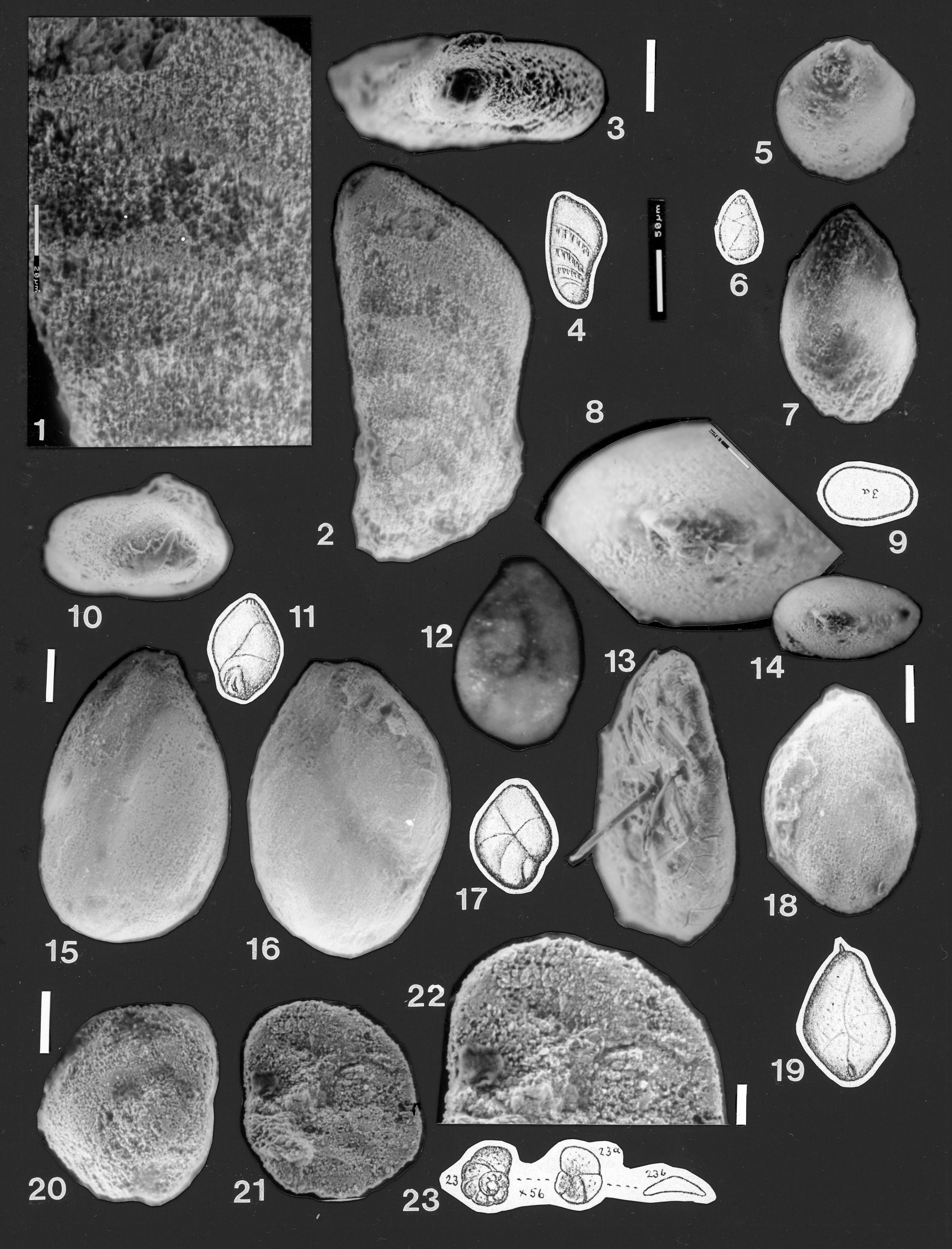
Fig. 6.1–4
1890 Flabellina dilatata Wisniowski, p. 230, plate 10, fig. 21.
1904 Flabellina dilatata Wisniowski; Chapman, p. 196, plate 22, fig. 18.
Material examined
Chapman’s single specimen, Museum of Victoria specimen P 2142.
Chapman’s remarks
From Chapman (1904, p. 196, for F. dilatata): “This species reminds one of the F. rugosa, from the Chalk of France, England and Bohemia, but it has the sutures practically flush with the surface of the test, whereas in F. rugosa they are salient.”
Description
Length ~0.325mm, width ~0.21mm, thickness variable, ~0.05–0.07mm. Test outline leaf-shaped, compressed, flat on lateral sides, periphery broadly rounded; relatively thick in relation to width and length. Left-hand side of specimen broken (see Fig. 6.1–2); Chapman’s drawing is apparently a reconstruction, unless the specimen has been broken since the drawing was made. Earliest chamber obscure; followed by two chambers in a linear vaginuline series, then an additional two chambers with a high chevron-shaped form. Sutures depressed but bordered along the basal margin of each chamber by a sharp ridge, which gives the appearance of thickened sutures, although the ridge is situated on the adjacent chamber wall anterior to the suture. Aperture terminal, approximately circular, poorly preserved. Wall calcareous, weathered and etched.
Subfamily Vaginulininae Reuss 1860
Genus Vaginulina d’Orbigny 1826
Vaginulina contracta (Terquem 1868)
Fig. 6.12–14
not 1826 Polymorphina burdigalensis d’Orbigny, p. 265, No. 2; Modèle No. 29.
not 1866 Cristellaria contracta Terquem, p. 440, pl. 18, figs. 9a–b.
1868 Marginulina contracta Terquem vars. A, B, F, G, H; Terquem, p. 125, 126, 127, plate 8, figs.14a–b, 15a–b, 19a–b, 20a–b, 21a–b; not vars. C, D, E.
1868 Marginulina solida Terquem var. D; Terquem, p. 123, pl. 8, figs. 5a, 5b.
1875 Cristellaria contracta Terquem and Berthelin, p. 42, pl. 3, figs. 15a–f (fide Ellis and Messina 1940 et seq.)
not 1882 Cristellaria contracta Terquem, p. 41, pl. 2, figs. 4, 5a–b.
1904 Polymorphina burdigalensis d’Orbigny 1826; Chapman, p.196–197, plate 22, fig. 19.
1937 Vaginulina legumen Linné (part); Bartenstein and Brand, p. 162, plate 15, fig. 10b only.
1937 ?Marginulina oolithica (Terquem) (part); Bartenstein and Brand, p. 160, plate 2A, fig. 11 only.
1955 ?Cristellaria polymorphinae Mityanina, p. 134, plate 3, fig. 1a–c (fide Ellis and Messina 1940 et seq.).
1965 Vaginulina contracta (Terquem); Hanzlikova, p. 86, plate 7, figs. 3a–b, 5a–b.
1982 Vaginulina flaccida Schwager; Coleman, plate 2, fig. 19.
Material examined
Chapman’s single specimen, Museum of Victoria specimen P 2143. Comparative material examined: Vaginulina flaccida Schwager of Coleman (1982, plate 2, fig. 19); specimen MPK 2414, British Geological Survey.
Chapman’s description
From Chapman (1904, p. 196, 197, for P. burdigalensis): “This species is distinguished by its compressed form, which, on one side at least, is decidedly concave; in this latter respect it is separable from P. compressa, D’Orbigny. The chambers are somewhat regularly alternate.”
Description
Test elongate, outline ovoid; shape moderately compressed, margins broadly rounded; test relatively thick (0.09mm) compared to length (0.32mm). Consists of three chambers in a uniserial, very slightly curved, astacoline or vaginuline arrangement. The initial chamber is slightly inflated, subtriangular in shape, and tapers obliquely to the dorsal margin. The two later chambers expand in width dorsally and ventrally, and have oblique, depressed sutures, which make an angle of 45˚ to the long axis of the test. The third chamber is moderately inflated, with the rounded aperture at the dorsal angle. The aperture is broken. The third chamber is deformed, with a groove running down the ventral margin of the final face.
Remarks
Chapman’s description and illustration of this specimen err, in that it is definitely not a polymorphinid, nor does it have alternating chambers. The second set of sutures drawn in Chapman’s figure (his Plate 22, figure 19) are merely surface artefacts - exfoliating chamber surface - due to the poor preservation of the specimen. The last chamber is slightly deformed, with a groove running down part of the ventral face (Fig. 6.12). The species is referred to Vaginulina contracta (Terquem 1868) because of the fairly rapid increase in chamber width (versus a fairly constant chamber height), the well-rounded periphery, and moderately thick test as seen in edge view.
Vaginulina cultrata (Terquem 1864)
Fig. 8.1–4
not 1863 Vaginulina schloenbachi Reuss, p. 46, pl. 3, fig. 6, (fide Ellis and Messina, 1940 et seq.)
1864 Marginulina cultrata Terquem, p. 411, pl. 9, fig. 11a–b (fide Ellis and Messina 1940 et seq.)
1904 Vaginulina schloenbachi Reuss var. interrupta Chapman, p. 190, pl. 22, fig. 5.
? 1957 Vaginulina toarica Mamontova, p. 185, pl. 3, fig. 1 (fide Ellis and Messina, 1940 et seq.)
1959 Vaginulina legumen (Linné); Cifelli, p. 322–323, pl. 5, figs. 15, 16.
1980 Vaginulina cf. listi (Bornemann); Coleman, pl. 4, fig. 19.
2001 Vaginulinopsis paracompressa Bartenstein and Malz (pars), p. 39, pl. 1, figs. 18, 19.
Material examined
Chapman’s single specimen, Museum of Victoria No. P2126.
Chapman’s description
From Chapman (1904, p. 190, for V. schloenbachi var. interrupta): “This variety is distinct from the specific form V. schloenbachi in having the long curved costae interrupted in the posterior area of each chamber. The costae are also more numerous, averaging six in each series in the variety, against three in the type form.”
Amended description
Length ~0.27mm, width ~0.06mm. Test medium-sized, compressed, consisting of >4 chambers in a linear arrangement. Lateral sides flat, periphery broadly rounded. Earliest chambers either broken off or obscured by adhering rock matrix. (Cleaning seemed inadvisable, due to the poor state of preservation of the wall). The test expands slowly in width and has a straight dorsal margin until the last chamber, on which the margin is slightly curved towards the ventral side. Sutures oblique, gently curved. Chamber height expands more on the dorsal margin than on the ventral margin, particularly in the first two chambers visible. The aperture is terminal, at the dorsal margin, a circular hole (blocked with debris) in an area of severely etched shell wall. Shell wall largely dissolved, non-costate on both sides, contrary to Chapman’s description. The sutures appear to have been flush with the chamber wall. This is inferred from the last chamber, the only area where the suture and wall are not largely dissolved.
The interrupted costate ornament which was described as the distinguishing characteristic of this subspecies is a weathering feature, caused by patchy dissolution of the shell wall (see Figure 8: 1). The wall is etched everywhere, but over the first three visible chambers the dissolution has caused deep, irregular pits penetrating in some places through the entire wall. Small remnant ridges of shell material adjacent to the sutures were mistaken by Chapman for ornament, but neither side of the specimen, in SEM or in reflected light at high magnification, shows any indication of ornament. For this reason, it is considered that the subspecies interrupta is not valid.
Remarks
The absence of ornament must argue against identification as Vaginulina schloenbachi Reuss. The impossibility of seeing the earliest chamber makes it unclear whether there is any initial coiling present, but a vaginuline chamber arrangement is tentatively suggested. The earliest name available for this Vaginulina appears to be Marginulina cultrata Terquem 1864, which may however lack the gently arched sutures seen on Chapman’s specimen. Vaginulina toarica Mamontova 1957 appears to be identical to the present specimen, but no view of the specimen in edge view is given. Vaginulina listi (Bornemann 1854) has a more curved dorsal margin and a slightly more rounded profile, with inflated rather than flat sides.
Vaginulina cf. protracta (Bornemann 1854)
Fig. 6.11, 15–18
not 1840 Marginulina compressa d’Orbigny, p. 17, pl. 1, figs. 18–19.
1854 Cristellaria protracta Bornemann, p. 39, pl. 4, figs. 27a–b.
not 1878 Cristellaria compressiuscula Marsson, new name (in error for M. compressa d’Orbigny, 1840).
1904 Marginulina compressa, D’Orbigny; Chapman, p. 188–189, pl. 22, fig. 4.
1936 Cristellaria (Astacolus) protracta Bornemann; Franke, p. 101, pl. 9, fig. 35.
Material examined
Chapman’s single specimen (Museum of Victoria specimen P2124).
Chapman’s remarks
From Chapman (1904, p. 188, 189, for his M. compressa): “The genera Marginulina and Vaginulina insensibly graduate into one another, exemplified in certain forms of the present type, the marginuline species becoming vaginuline by the continued compression of the test. Our specimen seems to occupy a place midway between these genera, but since it agrees with the original figure of Marginulina compressa given by D’Orbigny, rather than with the flatter type, Vaginulina legumen, Linné sp., it may be conveniently retained in the former genus.”
Description
Length ~1.15mm; width ~0.30mm; maximum thickness ~0.24mm. Test calcareous, elongate, moderately compressed. The large specimen is not well preserved but consists of probably five chambers arranged in a linear series, expanding slowly in width, without any visible initial enrolment. A sixth chamber may have been present (Figure 6: 11) but is now entirely broken away. The initial chamber has a blunt point at its base, and is not as rounded as Chapman’s drawing suggests (Figure 6: 17). The first three chambers are very difficult to see, in both the light microscope (Figure 6: 11) and ESEM, due to the flush sutures. These chambers are compressed and have a narrowly rounded margin both dorsally (Figure 6: 15) and ventrally (Figure 6: 16). The last two preserved chambers are better defined with strongly oblique, slightly depressed sutures. The final (preserved) chamber is more inflated, swollen ventrally (Figure 6: 16), but not much dorsally, the dorsal margin being still narrowly rounded as for the earlier part of the test. The aperture is at the tapering dorsal margin, and is somewhat produced, but broken off. The ventral surface of the last chamber is badly damaged, and small ridges suggest the presence of an additional chamber.
Remarks
This large specimen has initial chambers which appear to be arranged in a linear, Vaginulina-style, without an initial coil or curvature of the initial chambers. For this reason, Chapman’s identification as M. compressa d’Orbigny 1840 is not accepted, as this species is shown with a slightly curved initial portion, and is also described as slightly carinate. For the same reason, identification as Vaginulina listi (Bornemann 1854) is also rejected as Rabitz (1963) illustrated the holotype of V. listi as having the three initial chambers slightly enrolled. Vaginulina schraderensis Tappan 1960, from the Cretaceous, is shorter (0.74mm versus 1.15mm for Chapman’s specimen) but is similar in shape, particularly in the somewhat inflated final chamber.
Vaginulina solida (Terquem 1868)
Fig. 6.8–10
1868 Marginulina solida Terquem, 1868, p. 122, pl. 8, figs. 1–5.
1875 Cristellaria contracta Terquem and Berthelin, p. 42, pl. 3, fig. 15e only.
1886 Marginulina solida Terquem, p. 24, pl. 2, figs. 34–43.
?1890 Cristellaria debilis Wisniowski, p. 220, pl. 9, fig. 13.
1904 Marginulina solida Terquem; Chapman, p. 189, pl. 22, fig. 17.
1963 Astacolus debilis (Wisniowski); Espitalié and Sigal, p. 27, pl. 5, fig. 16.
2001 Vaginulina sp. A Bartenstein and Malz, p. 32, pl. 1, fig. 32; text-fig. 2.
Material examined
Chapman’s single specimen (Museum of Victoria specimen P2125).
Chapman’s remarks
From Chapman (1904, p. 189, for his M. solida): “The Australian specimen evidently belongs to this species [Marginulina solida Terquem, 1886], which, although variable, shows certain features which enable to place it in the group with Marginulina glabra, D’Orb., for a central type; this particular form being altogether slenderer and more compressed than the species just mentioned….”
Description
Length 0.27 mm; maximum thickness 0.07 mm. A small specimen, moderately compressed, composed of three chambers in a rectilinear arrangement, with very oblique sutures. Chamber arrangement not coiled, hence the assignment to Vaginulina. The initial chamber is more inflated and thicker in edge view than the second and third chambers. In side view the chambers expand in width, so that the test flares slightly. The margins of all chambers are broadly rounded. The two sutures are approximately parallel and are slightly depressed. The initial end of the first chamber is broken. The apertural area also has the wall broken away, and the aperture cannot be seen.
Remarks
Chapman placed his specimen in “Marginulina solida Terquem, 1886” and made no reference to the earlier use of the name by Terquem (1868). Chapman’s drawing of this specimen (his plate 22, figure 17, reproduced on Figure 6: 10 herein) is not accurate, and suggests an astacoline chamber arrangement. The arrangement of the chambers is more rectilinear and less coiled than his drawing suggests, and the second and third chambers do not taper as much along the ventral margin.
Vaginulina sp.
Fig. 6.5–7
not 1848 Marginulina lata Cornuel, p. 252, pl. 1, figs. 34–7.
not 1860 Marginulina lata Reuss, p. 206, pl. 5, fig. 7 (fide Ellis and Messina, 1940 et seq.)
1904 Vaginulina lata, Cornuel sp.; Chapman, p. 190, pl. 22, fig. 6.
Material examined
Chapman’s single specimen (Museum of Victoria specimen P2127).
Chapman’s remarks
From Chapman (1904, p. 190, 191, for his V. lata): “These compressed quasi cristellarian forms appear more properly to belong to the genus Vaginulina, as it is now understood, since the present type of shell has no distinct spiral commencement, and the test is uniformly flattened. The few-chambered, broad, compressed shells which may be referred to the above species are mostly characteristic of Mesozoic strata; but the species has also been recorded from recent soundings off Moncoeur Island, Bass Strait, at a depth of 38 fathoms. Our specimen somewhat resembles a closely allied form which was found in the Gault of France and England, Vaginulina biochei, Berthelin. The latter form, however, is distinguished by the thickened sutures of the chambers having a tendency to become salient, especially on the inner (anti-stoloniferous) margin of the test.”
Description
Length ~0.29 mm; width ~0.15 mm; thickness 0.07 mm. Broken specimen, possibly of two or three chambers, compressed, with broadly rounded margins. Surface essentially smooth, apart from surface etching; no chamber sutures visible. Aperture terminal, a broken hole. From the shape of the test the specimen is thought to be a Vaginulina, but no chamber outlines or sutures (apart from the broken, last suture at upper left) are visible, either in the light microscope or in the ESEM. Chapman’s drawing of the chambering (reproduced here as Fig. 6.5) appears to be based on irregular etched grooves due to weathering on the shell surface.
Genus Citharina d’Orbigny, 1839
Citharina sp. 1
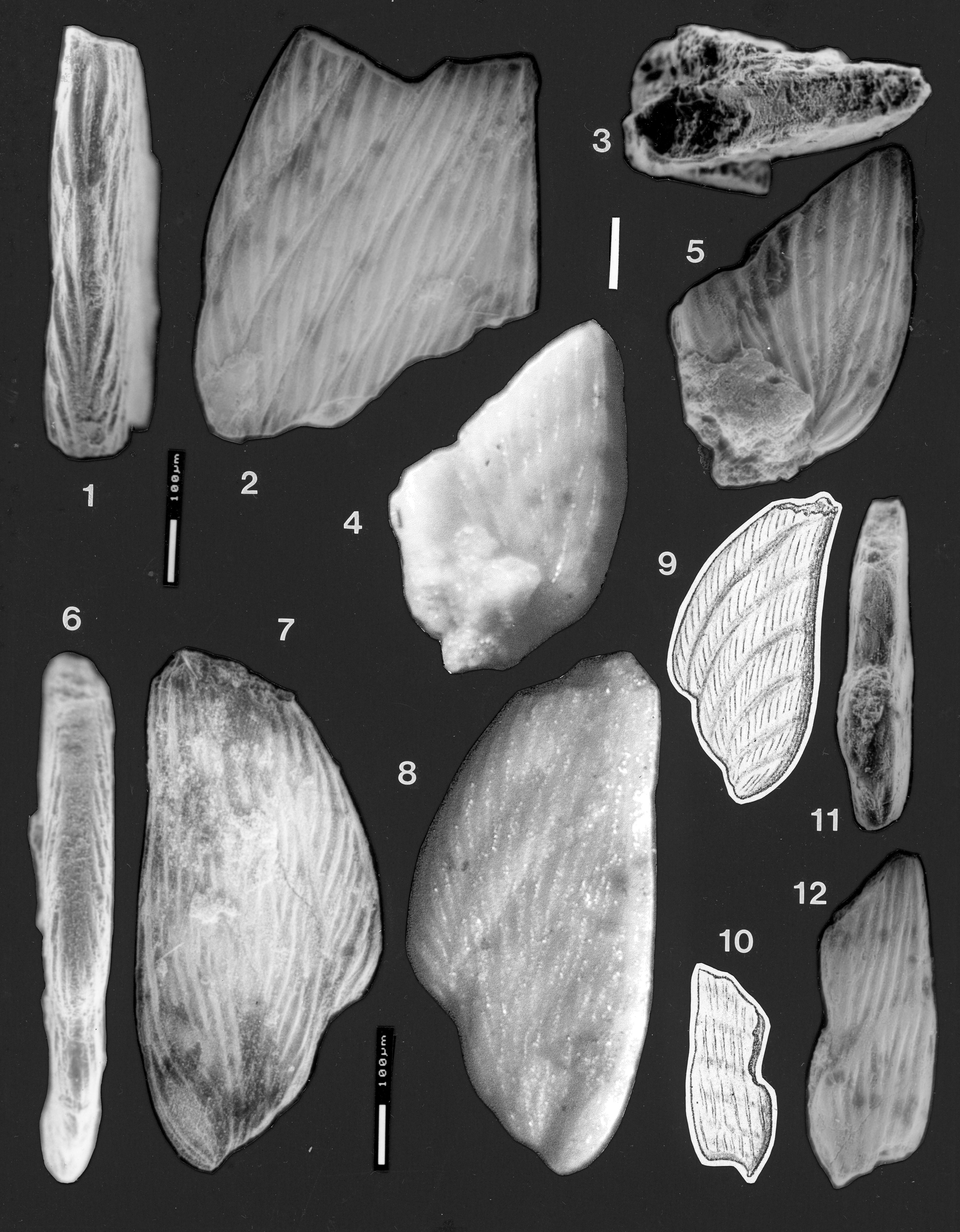
Fig. 7.3–5, 10–12
not 1846 Vaginulina (Citharina) strigillata Reuss, p. 106, pl. 24, fig. 29.
not 1864 Marginulina longuemari Terquem, var. clathrata Terquem, p. 192, pl. 8, figs. 16, 19a–b.
1904 Vaginulina strigillata Reuss; Chapman, p. 191, pl. 22, fig. 7 (P2128).
1904 Vaginulina intumescens Reuss (pars); Chapman, p. 191–2, (unfigured specimen P2131).
2001 Citharina clathrata (Terquem); Bartenstein & Malz, p. 33, pl. 2, figs. 44–46.
2001 ?Citharina subacuminata Bartenstein & Malz, p. 34, pl. 3, figs. 51–52.
Material examined
Chapman’s two specimens, Museum of Victoria specimens P 2128 (figured by Chapman) and P2131 (not figured by Chapman).
Chapman’s remarks on specimen P2128
From Chapman (1904, p. 191, for his V. strigillata): “…The particular form under notice is distinguished from others closely allied by the more pronounced striations on the surface of the shell, and which bridge over the suture lines, the latter being more or less depressed.”
Description of P2128
Length 0.56 mm; width 0.21 mm; thickness 0.07 mm. A fragment of a Citharina, broken at top and bottom, and possibly on the anterior edge, and abraded on all edges and on the ornament on the lateral sides. The raised costae are low but distinct, closely spaced (4-5 costae / ribs per 100 microns) on the lateral surfaces, slightly oblique to the length of the test, relatively straight, and mostly continuous across the sutures. Up to 8 ribs are present on the photographed side of the fragment (Fig. 7.12), which appears to be less than the true width of the original test. The costae become indistinct in places due possibly to abrasion of the higher parts of the sides, but appear to have been continuous. Some join together and disappear at the posterior margin. There is a slight suggestion of an abraded, low posterior keel.
Description of Chapman’s unfigured specimen P2131 (Fig. 7.3–5 herein)
Length >0.50 mm; width ~0.34 mm; thickness ~0.09 mm. Specimen consists of the initial and four following chambers; later chambers are broken away. The initial chamber may be oval but is partly obscured by matrix on both sides; the second and subsequent chambers expand in width rapidly, and very little in height, so that the test flares. The sides are flat; anterior margin is damaged; posterior margin has a very low keel. The sutures are only slightly depressed. There are about 12 costae of fairly uniform thickness, which radiate from the initial chamber as straight or occasionally bifurcating ridges, and which become oblique to the length of the test along the posterior margin. The costae continue essentially uninterrupted across the sutures (Fig. 7.5).
Comments
Chapman assigned specimen P2131 to Vaginulina intumescens, along with specimens P2129 and P2130 (see below). However, the presence of prominent, continuous, relatively straight or somewhat bifurcating costae (particularly as seen in the light microscope) suggests assignment of this fragment to the same species as P2128. The narrow shape of the latter is not considered significant, as the fragment appears to be broken, infilled and abraded along the anterior margin and the original width of the test is almost certainly not present. Due to the very incomplete nature of the specimens, no adequate identification can be suggested. The relative thickness in edge view and robustness of the tests does not suggest any affinity with the Cretaceous Citharina strigillata Reuss. The specimens cannot be compared to Citharina clathrata (Terquem 1864) as that species in the type specimen has only four longitudinal costae or ribs and is shown as sharply keeled on both margins. Terquem later (1868) defined different forms with many more striae and rounded anterior margins as Marginulina clathrata var. A, var. B and var. C. These resemble Chapman’s fragments in their elongate test form and numerous, straight and fairly continuous striae. However this name is a junior homonym, and the later forms would require renaming.
Citharina sp. 2
Fig. 7.1–2, 6–9
not 1863 Vaginulina intumescens Reuss, p. 49, pl. 4, fig. 2 (fide Ellis & Messina 1940 et seq.).
1868 ?Marginulina heteropleura Terquem, p. 116, pl. 7, figs. 19a–b.
1904 Vaginulina intumescens Reuss; Chapman, p. 191–192, pl. 22, fig. 8.
2001 ?Citharina geraldtonensis Bartenstein and Malz (part), p. 33, pl. 2, fig. 42 only, pl. 3, figs. 49–50.
Material examined
Chapman’s specimens P2129 (his Plate 22, figure 8) and P2130 (not illustrated by Chapman). Numerous specimens from Bringo Cutting locality, Geraldton area, Western Australia.
Chapman’s remarks
From Chapman (1904, p. 192, for his V. intumescens): “This species has, typically, a sub-triangular test, strongly compressed; ornamented with fine, curved, longitudinal striae. Terquem’s liassic specimen has more numerous and narrower chambers than ours.”
Description P2129
See Fig. 7.6–9, an almost complete specimen, length 0.85 mm, width 0.4 mm, thickness 0.09 mm. Test subtriangular in outline, flaring, laterally highly compressed with flat sides. Initial chamber elongated, ovoid, slightly inflated; followed with almost no curvature in the test axis, by six arcuate chambers. These expand rapidly in width, and slightly in height along the dorsal margin. Sutures gently curved, slightly depressed. Apertural region broken away. Sides ornamented with numerous wavey, somewhat discontinuous costae, variably parallel or slightly oblique to the length of the test, with a spacing of about 4 costae per 100 microns of wall. The costae are mostly interrupted at the sutures near the ventral margin but are more continuous close to the dorsal margin. The ridge along the sutures that is seen in specimen P2130 (see below) is developed to a small degree only on the final suture of specimen P2129 (see Fig. 7.7). The ventral margin is rounded but abraded (Fig. 7.6). The dorsal margin is sub-rounded, with a more prominent costa running along it.
Description P2130
See Fig. 7.1–2, a fragment, length >0.7 mm, width 0.562 mm, thickness 0.12 mm. A large fragment of a Citharina, consisting of more than five chambers from the middle portion of the test. Initial part (?half) of test missing, terminal part of last two chambers, including the aperture, also missing. Test large, sides flat with chambers oblique and sloping steeply back towards the initial part. Preserved chambers expand in height only slightly towards the dorsal margin. The sutures are slightly depressed. The most distinctive feature of this fragment is the variable form of the costae, which are oblique to the elongation of the test. In the last two chambers the costae taper and terminate at a low ridge along the suture; whilst in the earlier chambers the costae taper at the suture, bend parallel to the suture and run along it for some distance, then resume their former orientation and thickness across the next chamber. There is thus a ridge along the two latest sutures but not in the earlier sutures. The specimen thus has both “discontinuous” costae, and “continuous but undulating” costae, in close proximity. In ventral peripheral view (Fig. 7: 1), the fragment is highly compressed, with a narrowly rounded margin, surmounted by a short section of keel on the final chamber. On older chambers some costae wrap around the periphery and coalesce. On the dorsal margin a small but continuous keel is present on the part of the margin that is preserved.
Remarks
The variability both of test outline and costate ornament in Citharina presents great difficulties in defining the limits of species. Chapman’s specimens P2129 and P2130 appear similar to the holotype of Citharina geraldtonensis Bartenstein and Malz (2001), although Chapman’s specimen P2130 has perhaps heavier and more continuous costae. Chapman’s specimens do not closely resemble the paratypes of C. geraldtonensis, which have more closely spaced costae.
Family Polymorphinidae d’Orbigny 1839
Subfamily Polymorphininae d’Orbigny 1839
Genus Polymorphina d’Orbigny 1826
Polymorphina fontinensis Terquem 1874
Fig. 8.10–13, 15–17
not 1844 Polymorphina compressa Philippi, p. 69, pl. 1, fig. 35–35a.
not 1846 Polymorphina compressa d’Orbigny, p. 233, pl. 12, figs. 32–34.
not 1846 Polymorphina liassica Strickland, p. 31, text-figure b.
1874 Polymorphina fontinensis Terquem, p. 298, pl. 31, figs. 29–30.
1904 Polymorphina compressa d’Orbigny; Chapman, p. 197–198, pl. 22, figs. 21–22.
1959 Eoguttulina liassica (Strickland); Cifelli, p. 333, pl. 7, figs. 9–11.
1962 Eoguttulina liassica (Strickland); Lloyd, p. 370–372, pl. 1, figs. 1–3, text fig. 2A only.
1981 Eoguttulina liassica (Strickland); Mira and Martinez-Gallego, p. 326, pl. 1, fig. 10.
Material examined
Chapman’s specimens P2145 and P2146.
Chapman’s remarks
From Chapman (1904, p. 197, 198, for his P. compressa): “The Australian specimens are not quite typical, since they are much broader than D’Orbigny’s examples, and are sub-rhombic in outline; but the form is a very variable one, and it is perhaps better to refer it to this species rather than to regard it as a distinct form. In the prominence of the primordial chamber in at least one of our specimens it resembles P. bucculenta, Berthelin, from the Gault of France; it also shows a paucity of chambers and a similar arrangement as in that species, but the segments are not quite so strongly inflated”.
Description P2145
Length: 0.24 mm; width: 0.15 mm; thickness: 0.09 mm. Test elongate, tear-drop shaped, with a broadly rounded base; chambers compressed, with rounded margins (Fig. 8.10–11, 15). Composed of a poorly visible initial chamber, followed by three chambers, in an apparently biserial arrangement. Sutures shallowly depressed. Aperture terminal, at the tapering end of the final chamber. Opening of aperture obscured by heavy glue.
Description P2146
Length: 0.25 mm; width: 0.16 mm; thickness: not measurable. Test elongate, tear-drop shaped; composed of a faintly visible initial chamber at the rounded base, followed by two elongate chambers separated by a depressed suture (Fig. 8.12–13, 16–17). Aperture terminal, broken. The test is crushed and flattened, and on the reverse side is covered with fibres of cardboard from the original mount (Fig. 8.13). The specimen wall is cracked and disintegrating on this side.
Comments
Specimen P2145 is referred to Polymorphina (following Chapman) rather than Eoguttulina, on the basis of its weakly compressed, rather than circular, cross-section. Specimen P2146 is severely crushed and deformed and is tentatively referred to the same species as specimen P2145.
The specimens appear to be not conspecific with the Tertiary species Polymorphina compressa d’Orbigny 1846, because the latter is described as tapering at both extremities, whereas the initial parts of Chapman’s specimens are quite well rounded. (Polymorphina compressa d’Orbigny 1846 is a junior homonym of Polymorphina compressa Philippi 1844). The specimens appear to fall within the concept of “Eoguttulina” liassica (Strickland) of Lloyd (1962) and Mira and Martinez-Gallego (1981). This species does not appear to fit within Polymorphina liassica Strickland 1846, which was described as “pointed at both ends”, unlike the rounded initial part of Chapman’s specimens. Based on the rounded initial chamber, compressed profile with broadly rounded margins, and three-chambered shape, Chapman’s specimen P2145, and possibly also his P2146, are here referred to Polymorphina fontinensis Terquem 1874.
Polymorphina sp.
Fig. 8.8–9, 14, 18, 19
1904 Bulimina gregorii Chapman, p. 187–188, pl. 22, figs. 3, 3a.
Material examined
Chapman’s single specimen P2123.
Chapman’s description
From Chapman (1904, p. 187, 188, for his B. gregorii): “Test sub-rhomboidal, compressed, with rounded lateral edges. Aboral end terminated by a blunt spike. Aperture typically bulimine. Sutures faintly marked; chambers not very numerous. Shell-texture finely arenaceous. Length, 0.22mm, greatest width, 0.13mm. Affinities- The form of the test and comparative fewness of the chambers remind one of Bulimina pyrula, D’Orb. Our species, however, is readily distinguished from that form by the strong compression of the test. The two species B. caudigera, D’Orb. and B. ovula, D’Orb., also bear some relationship to our form, both in the compression of the test, and in having an aboral spine. The compression in our species is so marked, amounting to a concavity of the external surface, that it will be as well to regard it as a distinct form. It is worth noting that B. pyrula, D’Orb., occurs in certain jurassic clays in England, as recorded by Messrs. Jones and Parker (“triassic” clay of Chellaston). The above species is named after the Hon. A. C. Gregory, F.R.G.S., under whose auspices the larger fossils were collected, and who gave them to Mr. Daintree.”
Description
Length 0.17 mm, width 0.12 mm, thickness 0.07 mm. Test calcareous, ovoid, compressed oval profile with broadly rounded margins (Figure 8: 8, 14). Composed of a few chambers, tapering towards the apertural end; mode of coiling indeterminable; sutures not visible. Aperture terminal, slightly produced, obscured by debris. Wall calcareous, heavily etched.
Remarks
Chapman viewed and figured his species (Fig. 8.19) upside down compared to the orientation used here. Hence, he regarded the pointed end of the specimen as the initial end, and the fragment of rock about 10 microns high adhering to it as his “blunt spike” at the “aboral end”. He interpreted the “bulimine” aperture to lie in a slight depression at the broad end of the test, and a faint suture to begin at the aperture and extend towards the opposite end. The depression at Chapman’s aperture is less than he shows (Fig. 8.18–19). The wall in the vicinity is heavily etched, and there is a small dark hole that he interpreted as the aperture. Faint cracks and etching marks occur on the surface but are not as regular as Chapman’s drawing suggests. The author interprets this very badly preserved specimen to have an apical aperture (Fig. 8.8, 14), and based on this and its oval, compressed shape, assigns it to the genus Polymorphina d’Orbigny 1826. The absence of chambering information and the damaged aperture preclude assignment to any species.
Genus Pyrulina d’Orbigny 1839
Pyrulina gutta (d’Orbigny 1826)
Fig. 8.5–7
1826 Polymorphina (les Pyrulines) gutta d’Orbigny, p. 267, pl. 12, figs. 5–6.
1839 Pyrulina gutta (d’Orbigny), p. 267, 310.
1904 Polymorphina gutta d’Orbigny; Chapman, p. 197, Pl. 22, fig. 20.
1987 Pyrulina gutta (d’Orbigny); Loeblich and Tappan, p. 421, pl. 457, figs. 8–9.
Material examined
Chapman’s single specimen P2144.
Chapman’s remarks
From Chapman (1904, p. 197, for his P. gutta): “This is a well-known neocomian and gault species…One specimen.”
Description
Length ~0.16 mm; diameter ~ 0.08 mm. Test tear-drop shaped, with a rounded base and chambers tapering towards a terminal aperture. Cross-section almost circular. Test poorly preserved, with chamber arrangement indistinct, only one chamber (the final one) being visible, even when the specimen was wet. Sutures almost flush. The final chamber tapers towards a pointed aperture. Apertural form obscured by debris; possibly radiate.
Comment
The specimen appears to fit within d’Orbigny’s species but the absence of more than one visible chamber makes the identification tentative.
Suborder Robertinina Loeblich and Tappan 1984
Family ?Ceratobuliminidae Cushman 1927
Genus Lamarckella Kaptarenko-Chernousova 1956
? Lamarckella inflecta Kaptarenko-Chernousova 1956
Fig 8.20–23
1956 ? Lamarckella inflecta Kaptarenko-Chernousova, plate 1, fig. 5.
1959 ? Lamarckella inflecta Kaptarenko-Chernousova, plate 11, figs. 12–13.
1985 ? Paulina kurshensis Grigelis, plate 5, figs. 3, 4; plate 31, figs. 4–6; plate 32, figs. 1– 2.
not 1826 Rotalia (Rotalie) rosacea d’Orbigny, p. 273, mod`le no. 39, (fide Ellis & Messina, 1940 et seq.)
1904 Discorbina rosacea d’Orbigny sp.; Chapman, p. 198, pl. 22, figs. 23, 23a, b.
1937 Discorbis dreheri Bartenstein (part), p. 192, pl. 10, fig. 47c only.
not 1942 Asterigerina dreheri (Bartenstein) sensu ten Dam and Reinhold (=Reinholdella dreheri Brotzen 1948), p. 10–11, figs. 1–2.
not 1958 Discorbis lacunosus Antonova, p. 66, 67, pl. 4, figs.4a–c.
Material examined
Chapman’s single specimen P2147; Bartenstein and Brand (1937), specimens XXVII 666 a2—5, XXVII 666 a6—8; comparative specimens supplied by Dr. Bartenstein from the type area of Discorbis dreheri; ten Dam and Reinhold’s specimens F.410-1 and F.410-2 (= Reinholdella dreheri Brotzen 1948).
Chapman’s remarks
From Chapman (1904, p. 198, for his D. rosacea): “The occurrence of the above minute species is interesting as extending its range in time. It was previously recorded from the neocomian of England, besides being well-known as a tertiary fossil.”
Description
Length ~0.16mm; width ~0.14mm; height ~0.06mm. Test round to slightly oval in outline, slightly lobulate; trochospiral, a low convex dome on the spiral side, slightly concave on the umbilical side, with an acute periphery. On the spiral side, test consists of a proloculus followed by two whorls, each of about six or seven chambers, gently inflated, expanding slowly in size. Sutures oblique, directed backwards, slightly curved, flush to slightly depressed. Spiral suture a little deeper than the intercameral sutures. Chamber wall on this side heavily etched to wholly dissolved in places; some matrix adhering. On the umbilical side, shell material is present only on the earlier chambers; later chambers now consist only of an internal mould, previously with substantial matrix adhering to the last two chambers. Umbilical surface slightly concave, appearing to consist of six or seven chambers. Traces of sutures poorly preserved, slightly depressed, curved. Sutural trace of last chamber poorly preserved, marked by only a slight depression and indentation at the periphery, but with a re-entrant trough at right-angles to the suture. Second-last suture appearing more deeply incised, curved (Fig. 8.21–22). Final suture completely obscured by adhering sediment grains from the matrix.
._specimen_p2126._**57**_*pyrulina_gutta*_(dorbig.jpg)
Comments
The final chamber (most clearly visible in light microscope photos of the spiral side) is smaller than drawn by Chapman (Figure 8.23 herein, his figures 23, 23a, 23b). The removal of some matrix from the umbilical side has revealed more of the underlying mould, with a re-entrant in the surface of the last chamber. However, the preservation of the specimen is so poor that generic assignment remains questionable. The absence of an umbilical plug precludes assignment to Discorbis dreheri Bartenstein and Brand 1937 (Bartenstein, pers. comm., March 2001). Lamarckella inflecta Kaptarenko-Chernousova 1956 has a similar low planoconvex test shape with a depressed umbilical area, and of Jurassic species seems most similar in overall shape to Chapman’s poorly preserved specimen. Discorbis lacunosus Antonova 1958, from the Bajocian of the Caucasus, differs from Chapman’s specimen in having a much more rounded periphery and in being less convex on the spiral side.
ACKNOWLEDGMENTS
Dr Stefan Revets kindly borrowed Chapman’s specimens from the Museum of Victoria on behalf of the author, remounted, and photographed them in the ESEM™ (Environmental Scanning Electron Microscope), and without this substantial assistance, the present revision would not have been carried out. The University of Western Australia is thanked for providing electron microscope facilities. The Museum of Victoria is thanked for lending the specimens for study. Dr. Revets and Prof. David Haig are thanked for discussions on Jurassic taxonomy and stratigraphy. Ken Apthorpe is thanked for compiling Figure 1.